 Movie
Movie Controller


+ Open data

- Basic information
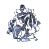
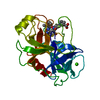
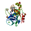
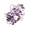
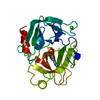
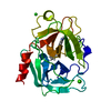
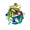
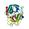
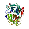
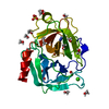
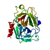
| Entry | Database: PDB / ID: 1qqu | ||||||
|---|---|---|---|---|---|---|---|
| Title | CRYSTAL STRUCTURE OF PORCINE BETA TRYPSIN WITH BOUND ACETATE ION | ||||||
 Components Components | BETA TRYPSIN | ||||||
 Keywords Keywords | HYDROLASE / SERINE PROTEASE | ||||||
| Function / homology |  Function and homology information Function and homology informationtrypsin / digestion / serine-type endopeptidase activity / proteolysis / : / metal ion binding Similarity search - Function | ||||||
| Biological species |  | ||||||
| Method |  X-RAY DIFFRACTION / MOLECULAR REPLCEMENT / Resolution: 1.63 Å X-RAY DIFFRACTION / MOLECULAR REPLCEMENT / Resolution: 1.63 Å | ||||||
 Authors Authors | Johnson, A. / Gautham, N. / Pattabhi, V. | ||||||
 Citation Citation | Journal: Biochim.Biophys.Acta / Year: 1999 Title: Crystal structure at 1.63 A resolution of the native form of porcine beta-trypsin: revealing an acetate ion binding site and functional water network. Authors: Johnson, A. / Gautham, N. / Pattabhi, V. #1: Journal: Acta Crystallogr.,Sect.D / Year: 1997Title: The First Crystal Structure at 1.8 Angstroms Resolution of an Active Autolysate Form of Porcine Alpha Trypsin Authors: Johnson, A. / Krishnaswamy, S. / Sundaram, P.V. / Pattabhi, V. | ||||||
| History |
|
- Structure visualization
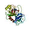
| Structure viewer | Molecule: MolmilJmol/JSmol |
|---|
- Downloads & links
-Download
| PDBx/mmCIF format | 1qqu.cif.gz | 58 KB | Display | PDBx/mmCIF format |
|---|---|---|---|---|
| PDB format | pdb1qqu.ent.gz | 41.3 KB | Display | PDB format |
| PDBx/mmJSON format | 1qqu.json.gz | Tree view | PDBx/mmJSON format | |
| Others |  Other downloads Other downloads |
-Validation report
| Arichive directory | https://data.pdbj.org/pub/pdb/validation_reports/qq/1qquftp://data.pdbj.org/pub/pdb/validation_reports/qq/1qqu | HTTPS FTP |
|---|
-Related structure data
| Related structure data | 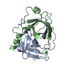 1aksS S: Starting model for refinement |
|---|---|
| Similar structure data |


-Links
 PDBj
PDBj

- Assembly
| Deposited unit | 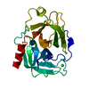
| ||||||||
|---|---|---|---|---|---|---|---|---|---|
| 1 |
| ||||||||
| Unit cell |
|
-Components
| #1: Protein | Mass: 23491.525 Da / Num. of mol.: 1 / Source method: isolated from a natural source / Source: (natural) |
|---|---|
| #2: Chemical | ChemComp-CA /   Mass: 40.078 Da / Num. of mol.: 1 / Source method: obtained synthetically / Formula: Ca Mass: 40.078 Da / Num. of mol.: 1 / Source method: obtained synthetically / Formula: Ca |
| #3: Chemical | ChemComp-ACT /   Mass: 59.044 Da / Num. of mol.: 1 / Source method: obtained synthetically / Formula: C2H3O2 Mass: 59.044 Da / Num. of mol.: 1 / Source method: obtained synthetically / Formula: C2H3O2 |
| #4: Water | ChemComp-HOH /  Mass: 18.015 Da / Num. of mol.: 149 / Source method: isolated from a natural source / Formula: H2O Mass: 18.015 Da / Num. of mol.: 149 / Source method: isolated from a natural source / Formula: H2O |
| Has protein modification | Y |
| Sequence details | THE 223 AMINO ACIDS OF TRYPSIN ARE IDENTIFIED |
-Experimental details
-Experiment
| Experiment | Method: X-RAY DIFFRACTION / Number of used crystals: 1 |
|---|
- Sample preparation
| Crystal | Density Matthews: 2.11 Å3/Da / Density % sol: 41.81 % | ||||||||||||||||||||||||||||||||||||||||||
|---|---|---|---|---|---|---|---|---|---|---|---|---|---|---|---|---|---|---|---|---|---|---|---|---|---|---|---|---|---|---|---|---|---|---|---|---|---|---|---|---|---|---|---|
| Crystal grow | pH: 6.7 / Details: pH 6.70 | ||||||||||||||||||||||||||||||||||||||||||
| Crystal grow | *PLUS Method: vapor diffusion, hanging dropDetails: drop consists of equal volume of protein and precipitant solutions | ||||||||||||||||||||||||||||||||||||||||||
| Components of the solutions | *PLUS
|
-Data collection
| Diffraction | Mean temperature: 293 K |
|---|---|
| Diffraction source | Wavelength: 1.54 |
| Detector | Type: MARRESEARCH / Detector: IMAGE PLATE / Date: Jan 13, 1997 |
| Radiation | Protocol: SINGLE WAVELENGTH / Monochromatic (M) / Laue (L): M / Scattering type: x-ray |
| Radiation wavelength | Wavelength: 1.54 Å / Relative weight: 1 |
| Reflection | Resolution: 1.63→23.62 Å / Num. obs: 21149 / % possible obs: 83.2 % / Observed criterion σ(I): 0 / Redundancy: 3.83 % / Rmerge(I) obs: 0.088 / Net I/σ(I): 19.96 |
| Reflection shell | Resolution: 1.63→1.7 Å / Redundancy: 3.2 % / Rmerge(I) obs: 0.4 / Mean I/σ(I) obs: 2.77 / % possible all: 80.6 |
| Reflection shell | *PLUS % possible obs: 80.6 % |
- Processing
| Software |
| ||||||||||||||||||||
|---|---|---|---|---|---|---|---|---|---|---|---|---|---|---|---|---|---|---|---|---|---|
| Refinement | Method to determine structure: MOLECULAR REPLCEMENT Starting model: 1AKS Resolution: 1.63→8 Å / Cross valid method: FREE R / σ(F): 0
| ||||||||||||||||||||
| Refinement step | Cycle: LAST / Resolution: 1.63→8 Å
| ||||||||||||||||||||
| Software | *PLUS Name: 'X-PLOR,REFMAC,CNS 0.9' / Classification: refinement | ||||||||||||||||||||
| Refine LS restraints | *PLUS
|
