 ムービー
ムービー コントローラー


+ データを開く

- 基本情報
基本情報
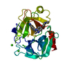
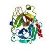
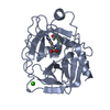
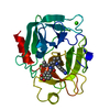
| 登録情報 | データベース: PDB / ID: 1qbn | ||||||
|---|---|---|---|---|---|---|---|
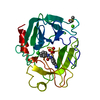
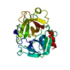
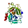
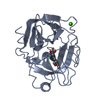
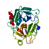
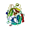
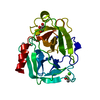
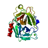
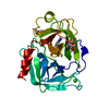
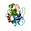
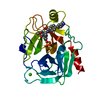
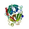
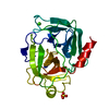
| タイトル | Bovine Trypsin 2-[amino(imino)methyl]-2-hydroxyphenoxy]-6-[3-(4,5-dihydro-1H-imidazol-2-yl)phenoxy]pyridine-4-carboxylic Acid (ZK-806688) Complex | ||||||
 要素 要素 | PROTEIN (TRYPSIN) | ||||||
 キーワード キーワード | HYDROLASE / SERINE PROTEINASE / PROTEIN-INHIBITOR COMPLEX / S1 POCKET | ||||||
| 機能・相同性 |  機能・相同性情報 機能・相同性情報trypsin / serpin family protein binding / serine protease inhibitor complex / digestion / endopeptidase activity / serine-type endopeptidase activity / proteolysis / : / metal ion binding 類似検索 - 分子機能 | ||||||
| 生物種 |  | ||||||
| 手法 |  X線回折 / DIRECT REPLACEMENT / 解像度: 1.8 Å X線回折 / DIRECT REPLACEMENT / 解像度: 1.8 Å | ||||||
 データ登録者 データ登録者 | Whitlow, M. | ||||||
 引用 引用 | ジャーナル: Acta Crystallogr.,Sect.D / 年: 1999 タイトル: Crystallographic analysis of potent and selective factor Xa inhibitors complexed to bovine trypsin. 著者: Whitlow, M. / Arnaiz, D.O. / Buckman, B.O. / Davey, D.D. / Griedel, B. / Guilford, W.J. / Koovakkat, S.K. / Liang, A. / Mohan, R. / Phillips, G.B. / Seto, M. / Shaw, K.J. / Xu, W. / Zhao, Z. ...著者: Whitlow, M. / Arnaiz, D.O. / Buckman, B.O. / Davey, D.D. / Griedel, B. / Guilford, W.J. / Koovakkat, S.K. / Liang, A. / Mohan, R. / Phillips, G.B. / Seto, M. / Shaw, K.J. / Xu, W. / Zhao, Z. / Light, D.R. / Morrissey, M.M. #1: ジャーナル: FEBS Lett. / 年: 1978タイトル: Crystal Structure Analysis and Refinement of Two Variants of Trigonal Trypsinogen 著者: Bode, W. / Huber, R. #2: ジャーナル: J.Med.Chem. / 年: 1999タイトル: Design, Synthesis, and Activity of 2,6-Diphenoxypyridine-Derived Factor Xa Inhibitors 著者: Phillips, G. / Davey, D. / Eagen, K. / Ng, H.P. / Pinkerton, M. / Koovakkat, S.K. / Whitlow, M. / Liang, A. / Trinh, L. / Morrissey, M.M. #3: ジャーナル: J.Med.Chem. / 年: 1998タイトル: Discovery of N-[2-[5-[Amino(imino)methyl]-2-hydroxyphenoxy]-3,5-difluoro-6-[3-(4,5-dihydro-1-methyl-1H-imidazol-2-yl)phenoxy] pyridin-4-yl-N-methylglycine (ZK-807834): A Potent, ...タイトル: Discovery of N-[2-[5-[Amino(imino)methyl]-2-hydroxyphenoxy]-3,5-difluoro-6-[3-(4,5-dihydro-1-methyl-1H-imidazol-2-yl)phenoxy] pyridin-4-yl-N-methylglycine (ZK-807834): A Potent, Selective and Orally Active Inhibitor of the Blood Coagulation Enzyme Factor Xa 著者: Phillips, G.B. / Buckman, B.O. / Davey, D.D. / Eagen, K.A. / Guilford, W.J. / Hinchman, J. / Ho, E. / Koovakkat, S. / Liang, A. / Light, D.R. / Mohan, R. / Ng, H.P. / Post, J. / Smith, D. / ...著者: Phillips, G.B. / Buckman, B.O. / Davey, D.D. / Eagen, K.A. / Guilford, W.J. / Hinchman, J. / Ho, E. / Koovakkat, S. / Liang, A. / Light, D.R. / Mohan, R. / Ng, H.P. / Post, J. / Smith, D. / Subramanyam, B. / Sullivan, M.E. / Trinh, L. / Vergona, R. / Walters, J. / White, K. / Whitlow, M. / Wu, S. / Xu, W. / Morrissey, M.M. #4: ジャーナル: FEBS Lett. / 年: 1995タイトル: Crystal structures of factor Xa specific inhibitors in complex with trypsin: structural grounds for inhibition of factor Xa and selectivity against thrombin 著者: Stubbs, M.T. / Huber, R. / Bode, W. #5: ジャーナル: Methods (San Diego) / 年: 1990タイトル: Analytical and Production Seeding Techniques 著者: Stura, E.A. / Wilson, I.A. #6: ジャーナル: J.Biol.Chem. / 年: 1996タイトル: X-ray structure of Active Site-inhibited Clotting Factor Xa 著者: Brandstetter, H. / Kuhne, A. / Bode, W. / Huber, R. / von der Saal, W. / Wirthensohn, K. / Engh, R.A. | ||||||
| 履歴 |
|
- 構造の表示
構造の表示
| 構造ビューア | 分子: MolmilJmol/JSmol |
|---|
- ダウンロードとリンク
-ダウンロード
| PDBx/mmCIF形式 | 1qbn.cif.gz | 60.7 KB | 表示 | PDBx/mmCIF形式 |
|---|---|---|---|---|
| PDB形式 | pdb1qbn.ent.gz | 43.3 KB | 表示 | PDB形式 |
| PDBx/mmJSON形式 | 1qbn.json.gz | ツリー表示 | PDBx/mmJSON形式 | |
| その他 |  その他のダウンロード その他のダウンロード |
-検証レポート
| アーカイブディレクトリ | https://data.pdbj.org/pub/pdb/validation_reports/qb/1qbnftp://data.pdbj.org/pub/pdb/validation_reports/qb/1qbn | HTTPS FTP |
|---|
-関連構造データ


-リンク
 PDBj
PDBj

- 集合体
集合体
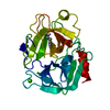
| 登録構造単位 | 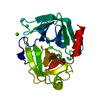
| ||||||||||
|---|---|---|---|---|---|---|---|---|---|---|---|
| 1 |
| ||||||||||
| 単位格子 |
| ||||||||||
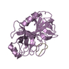
| 詳細 | The biological assembly is a monomer. |
-要素
| #1: タンパク質 | 分子量: 23324.287 Da / 分子数: 1 / 断片: BOVINE TRYPSIN / 由来タイプ: 天然 / 詳細: BOEHRINGER MANNHEIM CATALOG NUMBER 109,827 / 由来: (天然) | ||||||
|---|---|---|---|---|---|---|---|
| #2: 化合物 |   分子量: 40.078 Da / 分子数: 2 / 由来タイプ: 合成 / 式: Ca 分子量: 40.078 Da / 分子数: 2 / 由来タイプ: 合成 / 式: Ca#3: 化合物 | ChemComp-688 / |   分子量: 431.401 Da / 分子数: 1 / 由来タイプ: 合成 / 式: C22H17N5O5 分子量: 431.401 Da / 分子数: 1 / 由来タイプ: 合成 / 式: C22H17N5O5#4: 水 | ChemComp-HOH / |  分子量: 18.015 Da / 分子数: 163 / 由来タイプ: 天然 / 式: H2O 分子量: 18.015 Da / 分子数: 163 / 由来タイプ: 天然 / 式: H2OHas protein modification | Y | |
-実験情報
-実験
| 実験 | 手法: X線回折 / 使用した結晶の数: 1 |
|---|
- 試料調製
試料調製
| 結晶 | マシュー密度: 2.05 Å3/Da / 溶媒含有率: 39.82 % 解説: STARTING FROM THE TRYPSIN 2-AMINOBENZIMIDAZOLE STRUCTURE | ||||||||||||||||||||||||||||||||||||
|---|---|---|---|---|---|---|---|---|---|---|---|---|---|---|---|---|---|---|---|---|---|---|---|---|---|---|---|---|---|---|---|---|---|---|---|---|---|
| 結晶化 | 温度: 297 K / 手法: 蒸気拡散法, ハンギングドロップ法 / pH: 7.4 詳細: MICROCRYSTALS GROWN USING THE HANGING DROP METHOD IN LINBRO CULTURE PLATES. CRYSTALLIZATION RESERVOIRS CONTAIN 1.8 - 2.0 M AMMONIUM SULFATE, 50 MM TRIS HCL, 10 MM CACL2, PH 7.4. SIX UL OF 15 ...詳細: MICROCRYSTALS GROWN USING THE HANGING DROP METHOD IN LINBRO CULTURE PLATES. CRYSTALLIZATION RESERVOIRS CONTAIN 1.8 - 2.0 M AMMONIUM SULFATE, 50 MM TRIS HCL, 10 MM CACL2, PH 7.4. SIX UL OF 15 MG/ML BOVINE TRYPSIN IN 20 MM 2-AMINOBENZIMIDAZOLE WAS ADDED TO 6 UL OF RESERVOIR. MACROSEEDING WAS USED TO GROW LARGE CRYSTALS (REF 5). SINGLE CRYSTALS WERE WASHED IN 0.8 M AMMONIUM SULFATE, 50 MM TRIS HCL, 10 MM CACL2, PH 7.4. THE WASHED CRYSTALS WERE PLACED IN A 18 HOUR OLD HANGING, VAPOR DIFFUSION, HANGING DROP, temperature 297K | ||||||||||||||||||||||||||||||||||||
| 結晶化 | *PLUS | ||||||||||||||||||||||||||||||||||||
| 溶液の組成 | *PLUS
|
-データ収集
| 回折 | 平均測定温度: 297 K |
|---|---|
| 放射光源 | 由来: 回転陽極 / タイプ: RIGAKU RU300 / 波長: 1.5418 |
| 検出器 | タイプ: RIGAKU RAXIS IIC / 検出器: IMAGE PLATE / 日付: 1994年10月15日 / 詳細: MIRRORS |
| 放射 | プロトコル: SINGLE WAVELENGTH / 単色(M)・ラウエ(L): M / 散乱光タイプ: x-ray |
| 放射波長 | 波長: 1.5418 Å / 相対比: 1 |
| 反射 | 解像度: 1.8→10 Å / Num. all: 16885 / Num. obs: 16575 / % possible obs: 95.2 % / Observed criterion σ(I): 2 / 冗長度: 7.4 % / Rsym value: 0.075 / Net I/σ(I): 9.7 |
| 反射 シェル | 解像度: 1.8→1.9 Å / 冗長度: 5.1 % / Mean I/σ(I) obs: 2.36 / Rsym value: 0.226 / % possible all: 90 |
| 反射 | *PLUS Rmerge(I) obs: 0.075 |
| 反射 シェル | *PLUS 最高解像度: 1.8 Å / 最低解像度: 1.9 Å / % possible obs: 90 % / Rmerge(I) obs: 0.226 |
- 解析
解析
| ソフトウェア |
| ||||||||||||||||||||||||||||||||||||||||||||||||||||||||||||||||||||||||||||||||||||
|---|---|---|---|---|---|---|---|---|---|---|---|---|---|---|---|---|---|---|---|---|---|---|---|---|---|---|---|---|---|---|---|---|---|---|---|---|---|---|---|---|---|---|---|---|---|---|---|---|---|---|---|---|---|---|---|---|---|---|---|---|---|---|---|---|---|---|---|---|---|---|---|---|---|---|---|---|---|---|---|---|---|---|---|---|---|
| 精密化 | 構造決定の手法: DIRECT REPLACEMENT / 解像度: 1.8→10 Å / 交差検証法: THROUGHOUT / σ(F): 2 立体化学のターゲット値: Hendrickson, W.A. (1985): Methods in Enzymology 115, pp. 252-270
| ||||||||||||||||||||||||||||||||||||||||||||||||||||||||||||||||||||||||||||||||||||
| 精密化ステップ | サイクル: LAST / 解像度: 1.8→10 Å
| ||||||||||||||||||||||||||||||||||||||||||||||||||||||||||||||||||||||||||||||||||||
| 拘束条件 |
| ||||||||||||||||||||||||||||||||||||||||||||||||||||||||||||||||||||||||||||||||||||
| ソフトウェア | *PLUS 名称: PROFFT / 分類: refinement | ||||||||||||||||||||||||||||||||||||||||||||||||||||||||||||||||||||||||||||||||||||
| 精密化 | *PLUS % reflection Rfree: 4 % / Rfactor obs: 0.154 / Rfactor Rwork: 0.155 | ||||||||||||||||||||||||||||||||||||||||||||||||||||||||||||||||||||||||||||||||||||
| 溶媒の処理 | *PLUS | ||||||||||||||||||||||||||||||||||||||||||||||||||||||||||||||||||||||||||||||||||||
| 原子変位パラメータ | *PLUS | ||||||||||||||||||||||||||||||||||||||||||||||||||||||||||||||||||||||||||||||||||||
| 拘束条件 | *PLUS
|
