 Movie
Movie Controller


+ Open data

- Basic information
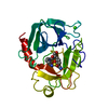
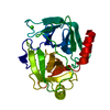
| Entry | Database: PDB / ID: 1qa0 | |||||||||
|---|---|---|---|---|---|---|---|---|---|---|
| Title | BOVINE TRYPSIN 2-AMINOBENZIMIDAZOLE COMPLEX | |||||||||
 Components Components | TRYPSIN | |||||||||
 Keywords Keywords | HYDROLASE / PROTEIN-INHIBITOR COMPLEX / S1 POCKET / SERINE PROTEASE | |||||||||
| Function / homology |  Function and homology information Function and homology informationtrypsin / serpin family protein binding / serine protease inhibitor complex / digestion / endopeptidase activity / serine-type endopeptidase activity / proteolysis / : / metal ion binding Similarity search - Function | |||||||||
| Biological species |  | |||||||||
| Method |  X-RAY DIFFRACTION / Resolution: 1.8 Å X-RAY DIFFRACTION / Resolution: 1.8 Å | |||||||||
 Authors Authors | Whitlow, M. | |||||||||
 Citation Citation | Journal: Acta Crystallogr.,Sect.D / Year: 1999 Title: Crystallographic analysis of potent and selective factor Xa inhibitors complexed to bovine trypsin. Authors: Whitlow, M. / Arnaiz, D.O. / Buckman, B.O. / Davey, D.D. / Griedel, B. / Guilford, W.J. / Koovakkat, S.K. / Liang, A. / Mohan, R. / Phillips, G.B. / Seto, M. / Shaw, K.J. / Xu, W. / Zhao, Z. ...Authors: Whitlow, M. / Arnaiz, D.O. / Buckman, B.O. / Davey, D.D. / Griedel, B. / Guilford, W.J. / Koovakkat, S.K. / Liang, A. / Mohan, R. / Phillips, G.B. / Seto, M. / Shaw, K.J. / Xu, W. / Zhao, Z. / Light, D.R. / Morrissey, M.M. #1: Journal: FEBS Lett. / Year: 1978Title: Crystal Structure Analysis and Refinement of Two Variants of Trigonal Trypsinogen Authors: Bode, W. / Huber, R. #2: Journal: Methods (San Diego) / Year: 1990Title: ANALYTICAL AND PRODUCTION SEEDING TECHNIQUES Authors: Stura, E.A. / Wilson, I.A. | |||||||||
| History |
|
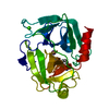
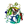
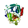
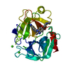
- Structure visualization
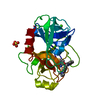
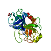
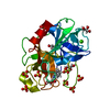
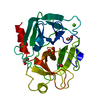
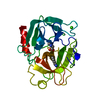
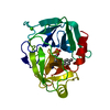
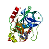
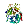
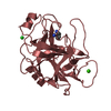
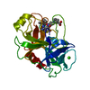
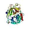
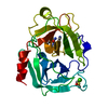
| Structure viewer | Molecule: MolmilJmol/JSmol |
|---|
- Downloads & links
-Download
| PDBx/mmCIF format | 1qa0.cif.gz | 58.1 KB | Display | PDBx/mmCIF format |
|---|---|---|---|---|
| PDB format | pdb1qa0.ent.gz | 40.8 KB | Display | PDB format |
| PDBx/mmJSON format | 1qa0.json.gz | Tree view | PDBx/mmJSON format | |
| Others |  Other downloads Other downloads |
-Validation report
| Arichive directory | https://data.pdbj.org/pub/pdb/validation_reports/qa/1qa0ftp://data.pdbj.org/pub/pdb/validation_reports/qa/1qa0 | HTTPS FTP |
|---|
-Related structure data


-Links
 PDBj
PDBj

- Assembly
| Deposited unit | 
| ||||||||||
|---|---|---|---|---|---|---|---|---|---|---|---|
| 1 |
| ||||||||||
| Unit cell |
|
-Components
| #1: Protein | Mass: 23324.287 Da / Num. of mol.: 1 / Fragment: BOVINE TRYPSIN / Mutation: NONE / Source method: isolated from a natural source / Source: (natural) |
|---|---|
| #2: Chemical | ChemComp-CA /   Mass: 40.078 Da / Num. of mol.: 1 / Source method: obtained synthetically / Formula: Ca / Details: BOEHRINGER MANNHEIM CORP. 109,827, ALRICH 17,177-8 Mass: 40.078 Da / Num. of mol.: 1 / Source method: obtained synthetically / Formula: Ca / Details: BOEHRINGER MANNHEIM CORP. 109,827, ALRICH 17,177-8 |
| #3: Chemical | ChemComp-AX7 / 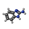  Mass: 133.151 Da / Num. of mol.: 1 / Source method: obtained synthetically / Formula: C7H7N3 / Details: SIGMA C 5080 Mass: 133.151 Da / Num. of mol.: 1 / Source method: obtained synthetically / Formula: C7H7N3 / Details: SIGMA C 5080 |
| #4: Water | ChemComp-HOH /  Mass: 18.015 Da / Num. of mol.: 125 / Source method: isolated from a natural source / Formula: H2O Mass: 18.015 Da / Num. of mol.: 125 / Source method: isolated from a natural source / Formula: H2O |
| Has protein modification | Y |
-Experimental details
-Experiment
| Experiment | Method: X-RAY DIFFRACTION / Number of used crystals: 1 |
|---|
- Sample preparation
| Crystal | Density Matthews: 2.04 Å3/Da / Density % sol: 39.82 % | ||||||||||||||||||||||||||||||||||||
|---|---|---|---|---|---|---|---|---|---|---|---|---|---|---|---|---|---|---|---|---|---|---|---|---|---|---|---|---|---|---|---|---|---|---|---|---|---|
| Crystal grow | Temperature: 298 K / Method: vapor diffusion, hanging drop / pH: 7.4 Details: MICROCRYSTALS WERE INITIALLY GROWN USING THE HANGING DROP METHOD IN LINBRO CULTURE PLATES. CRYSTALLIZATION RESERVOIRS CONTAIN 1.8 - 2.0 M AMMONIUM SULFATE, 50 MM TRIS HCL, 10 MM CACL2, PH 7. ...Details: MICROCRYSTALS WERE INITIALLY GROWN USING THE HANGING DROP METHOD IN LINBRO CULTURE PLATES. CRYSTALLIZATION RESERVOIRS CONTAIN 1.8 - 2.0 M AMMONIUM SULFATE, 50 MM TRIS HCL, 10 MM CACL2, PH 7.4. SIX UL OF 15 MG/ML BOVINE TRYPSIN IN 20 MM 2-AMINOBENZIMIDAZOLE WAS ADDED TO 6 UL OF RESERVOIR. MACROSEEDING WAS USED TO GROW LARGE CRYSTALS (REF 2). SINGLE CRYSTALS WERE WASHED IN 0.8 M AMMONIUM SULFATE, 50 MM TRIS HCL, 10 MM CACL2, PH 7.4. THE WASHED CRYSTALS WERE PLACED IN A 18 HOUR OLD HANGING., VAPOR DIFFUSION, HANGING DROP, temperature 298K | ||||||||||||||||||||||||||||||||||||
| Crystal grow | *PLUS | ||||||||||||||||||||||||||||||||||||
| Components of the solutions | *PLUS
|
-Data collection
| Diffraction | Mean temperature: 298 K |
|---|---|
| Diffraction source | Source: ROTATING ANODE / Type: RIGAKU RU300 / Wavelength: 1.5418 |
| Detector | Type: RIGAKU RAXIS II / Detector: IMAGE PLATE / Date: Oct 10, 1994 |
| Radiation | Protocol: SINGLE WAVELENGTH / Monochromatic (M) / Laue (L): M / Scattering type: x-ray |
| Radiation wavelength | Wavelength: 1.5418 Å / Relative weight: 1 |
| Reflection | Resolution: 1.8→10 Å / Num. all: 16893 / Num. obs: 16893 / % possible obs: 91.1 % / Observed criterion σ(F): 0 / Observed criterion σ(I): 0 / Redundancy: 6.3 % / Rmerge(I) obs: 0.095 / Net I/σ(I): 8.1 |
| Reflection shell | Resolution: 1.8→1.9 Å / Redundancy: 4 % / Rmerge(I) obs: 0.022 / % possible all: 78.5 |
| Reflection | *PLUS |
| Reflection shell | *PLUS Highest resolution: 1.8 Å / Lowest resolution: 1.9 Å / % possible obs: 78.5 % / Rmerge(I) obs: 0.0262 / Mean I/σ(I) obs: 2.2 |
- Processing
| Software |
| ||||||||||||||||||||||||||||||||||||||||||||||||||||||||||||||||||||||||||||||||||||
|---|---|---|---|---|---|---|---|---|---|---|---|---|---|---|---|---|---|---|---|---|---|---|---|---|---|---|---|---|---|---|---|---|---|---|---|---|---|---|---|---|---|---|---|---|---|---|---|---|---|---|---|---|---|---|---|---|---|---|---|---|---|---|---|---|---|---|---|---|---|---|---|---|---|---|---|---|---|---|---|---|---|---|---|---|---|
| Refinement | Resolution: 1.8→10 Å / σ(F): 2 / Details: USED RESTRAINED LEAST SQUARES /
| ||||||||||||||||||||||||||||||||||||||||||||||||||||||||||||||||||||||||||||||||||||
| Refinement step | Cycle: LAST / Resolution: 1.8→10 Å
| ||||||||||||||||||||||||||||||||||||||||||||||||||||||||||||||||||||||||||||||||||||
| Refine LS restraints |
| ||||||||||||||||||||||||||||||||||||||||||||||||||||||||||||||||||||||||||||||||||||
| Software | *PLUS Name: PROFFT / Classification: refinement | ||||||||||||||||||||||||||||||||||||||||||||||||||||||||||||||||||||||||||||||||||||
| Refinement | *PLUS Highest resolution: 1.8 Å / σ(F): 2 | ||||||||||||||||||||||||||||||||||||||||||||||||||||||||||||||||||||||||||||||||||||
| Solvent computation | *PLUS | ||||||||||||||||||||||||||||||||||||||||||||||||||||||||||||||||||||||||||||||||||||
| Displacement parameters | *PLUS | ||||||||||||||||||||||||||||||||||||||||||||||||||||||||||||||||||||||||||||||||||||
| Refine LS restraints | *PLUS
|
