 Movie
Movie Controller


+ Open data

- Basic information
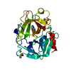
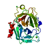
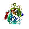
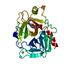
| Entry | Database: PDB / ID: 1auj | ||||||
|---|---|---|---|---|---|---|---|
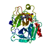
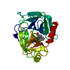
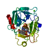
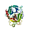
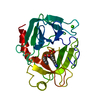
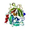
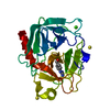
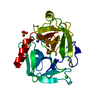
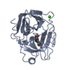
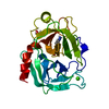
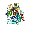
| Title | BOVINE TRYPSIN COMPLEXED TO META-CYANO-BENZYLIC INHIBITOR | ||||||
 Components Components | TRYPSIN | ||||||
 Keywords Keywords | HYDROLASE / SERINE PROTEINASE | ||||||
| Function / homology |  Function and homology information Function and homology informationtrypsin / serpin family protein binding / serine protease inhibitor complex / digestion / endopeptidase activity / serine-type endopeptidase activity / proteolysis / : / metal ion binding Similarity search - Function | ||||||
| Biological species |  | ||||||
| Method |  X-RAY DIFFRACTION / DIFFERENCE / Resolution: 2.1 Å X-RAY DIFFRACTION / DIFFERENCE / Resolution: 2.1 Å | ||||||
 Authors Authors | Alexander, R. / Smallwood, A. / Kettner, C. | ||||||
 Citation Citation | Journal: Biochemistry / Year: 1997 Title: New inhibitors of thrombin and other trypsin-like proteases: hydrogen bonding of an aromatic cyano group with a backbone amide of the P1 binding site replaces binding of a basic side chain. Authors: Lee, S.L. / Alexander, R.S. / Smallwood, A. / Trievel, R. / Mersinger, L. / Weber, P.C. / Kettner, C. #1: Journal: Biochemistry / Year: 1995Title: Kinetic and Crystallographic Studies of Thrombin with Ac-(D)Phe-Pro-Boroarg-Oh and its Lysine, Amidine, Homolysine, and Ornithine Analogs Authors: Weber, P.C. / Lee, S.L. / Lewandowski, F.A. / Schadt, M.C. / Chang, C.W. / Kettner, C.A. #2: Journal: FEBS Lett. / Year: 1975Title: The Single Calcium-Binding Site of Crystallin Bovin Beta-Trypsin Authors: Bode, W. / Schwager, P. #3: Journal: J.Mol.Biol. / Year: 1975Title: The Refined Crystal Structure of Bovine Beta-Trypsin at 1.8 A Resolution. I. Crystallization, Data Collection and Application of Patterson Search Technique Authors: Fehlhammer, H. / Bode, W. | ||||||
| History |
|
- Structure visualization
| Structure viewer | Molecule: MolmilJmol/JSmol |
|---|
- Downloads & links
-Download
| PDBx/mmCIF format | 1auj.cif.gz | 58.1 KB | Display | PDBx/mmCIF format |
|---|---|---|---|---|
| PDB format | pdb1auj.ent.gz | 41.2 KB | Display | PDB format |
| PDBx/mmJSON format | 1auj.json.gz | Tree view | PDBx/mmJSON format | |
| Others |  Other downloads Other downloads |
-Validation report
| Arichive directory | https://data.pdbj.org/pub/pdb/validation_reports/au/1aujftp://data.pdbj.org/pub/pdb/validation_reports/au/1auj | HTTPS FTP |
|---|
-Related structure data
| Similar structure data |
|---|


-Links
 PDBj
PDBj

- Assembly
| Deposited unit | 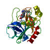
| ||||||||
|---|---|---|---|---|---|---|---|---|---|
| 1 |
| ||||||||
| Unit cell |
|
-Components
| #1: Protein | Mass: 23324.287 Da / Num. of mol.: 1 / Source method: isolated from a natural source / Source: (natural) |
|---|---|
| #2: Chemical | ChemComp-CA /   Mass: 40.078 Da / Num. of mol.: 1 / Source method: obtained synthetically / Formula: Ca Mass: 40.078 Da / Num. of mol.: 1 / Source method: obtained synthetically / Formula: Ca |
| #3: Chemical | ChemComp-PPB /   Mass: 434.296 Da / Num. of mol.: 1 / Source method: obtained synthetically / Formula: C23H27BN4O4 Mass: 434.296 Da / Num. of mol.: 1 / Source method: obtained synthetically / Formula: C23H27BN4O4 |
| #4: Water | ChemComp-HOH /  Mass: 18.015 Da / Num. of mol.: 140 / Source method: isolated from a natural source / Formula: H2O Mass: 18.015 Da / Num. of mol.: 140 / Source method: isolated from a natural source / Formula: H2O |
| Has protein modification | Y |
-Experimental details
-Experiment
| Experiment | Method: X-RAY DIFFRACTION / Number of used crystals: 1 |
|---|
- Sample preparation
| Crystal | Density Matthews: 2.33 Å3/Da / Density % sol: 47 % | ||||||||||||||||||
|---|---|---|---|---|---|---|---|---|---|---|---|---|---|---|---|---|---|---|---|
| Crystal grow | pH: 6.6 / Details: pH 6.6 | ||||||||||||||||||
| Crystal | *PLUS | ||||||||||||||||||
| Crystal grow | *PLUS pH: 7.5 / Method: batch method / Details: Krieger, M., (1974) J. Mol. Biol., 83, 209. | ||||||||||||||||||
| Components of the solutions | *PLUS
|
-Data collection
| Diffraction | Mean temperature: 273 K |
|---|---|
| Diffraction source | Source: ROTATING ANODE / Type: RIGAKU RUH2R / Wavelength: 1.5418 |
| Detector | Type: RIGAKU / Detector: IMAGE PLATE / Date: Sep 1, 1995 |
| Radiation | Monochromator: NI FILTER / Monochromatic (M) / Laue (L): M / Scattering type: x-ray |
| Radiation wavelength | Wavelength: 1.5418 Å / Relative weight: 1 |
| Reflection | Highest resolution: 2.1 Å / % possible obs: 90 % / Observed criterion σ(I): 1 / Rsym value: 0.052 |
| Reflection | *PLUS Rmerge(I) obs: 0.052 |
- Processing
| Software |
| ||||||||||||||||||||||||||||||||||||||||||||||||||||||||||||
|---|---|---|---|---|---|---|---|---|---|---|---|---|---|---|---|---|---|---|---|---|---|---|---|---|---|---|---|---|---|---|---|---|---|---|---|---|---|---|---|---|---|---|---|---|---|---|---|---|---|---|---|---|---|---|---|---|---|---|---|---|---|
| Refinement | Method to determine structure: DIFFERENCE / Resolution: 2.1→8 Å / σ(F): 1 /
| ||||||||||||||||||||||||||||||||||||||||||||||||||||||||||||
| Refinement step | Cycle: LAST / Resolution: 2.1→8 Å
| ||||||||||||||||||||||||||||||||||||||||||||||||||||||||||||
| Refine LS restraints |
|
