 Movie
Movie Controller

[English] 日本語
 Yorodumi
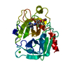
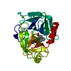
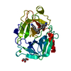
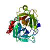
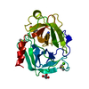
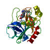
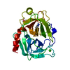
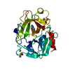
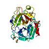
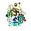
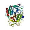
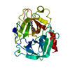
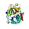
- PDB-3uqv: Bovine trypsin variant X(triplePhe227) in complex with small mole... -
+ Open data

- Basic information
| Entry | Database: PDB / ID: 3uqv | ||||||
|---|---|---|---|---|---|---|---|
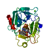
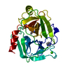
| Title | Bovine trypsin variant X(triplePhe227) in complex with small molecule inhibitor | ||||||
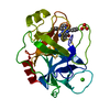
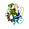
 Components Components | Cationic trypsin | ||||||
 Keywords Keywords | HYDROLASE/HYDROLASE inhibitor / Trypsin-like serine protease / HYDROLASE / PROTEIN BINDING / DUODENUM / HYDROLASE-HYDROLASE inhibitor complex | ||||||
| Function / homology |  Function and homology information Function and homology informationtrypsin / serpin family protein binding / serine protease inhibitor complex / digestion / endopeptidase activity / serine-type endopeptidase activity / proteolysis / : / metal ion binding Similarity search - Function | ||||||
| Biological species |  | ||||||
| Method |  X-RAY DIFFRACTION / MOLECULAR REPLACEMENT / Resolution: 2.4 Å X-RAY DIFFRACTION / MOLECULAR REPLACEMENT / Resolution: 2.4 Å | ||||||
 Authors Authors | Tziridis, A. / Neumann, P. / Kolenko, P. / Stubbs, M.T. | ||||||
 Citation Citation | Journal: Biol.Chem. / Year: 2014 Title: Correlating structure and ligand affinity in drug discovery: a cautionary tale involving second shell residues. Authors: Tziridis, A. / Rauh, D. / Neumann, P. / Kolenko, P. / Menzel, A. / Brauer, U. / Ursel, C. / Steinmetzer, P. / Sturzebecher, J. / Schweinitz, A. / Steinmetzer, T. / Stubbs, M.T. | ||||||
| History |
|
- Structure visualization
| Structure viewer | Molecule: MolmilJmol/JSmol |
|---|
- Downloads & links
-Download
| PDBx/mmCIF format | 3uqv.cif.gz | 61.5 KB | Display | PDBx/mmCIF format |
|---|---|---|---|---|
| PDB format | pdb3uqv.ent.gz | 43.1 KB | Display | PDB format |
| PDBx/mmJSON format | 3uqv.json.gz | Tree view | PDBx/mmJSON format | |
| Others |  Other downloads Other downloads |
-Validation report
| Arichive directory | https://data.pdbj.org/pub/pdb/validation_reports/uq/3uqvftp://data.pdbj.org/pub/pdb/validation_reports/uq/3uqv | HTTPS FTP |
|---|
-Related structure data
| Related structure data | 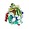 3plbC 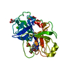 3plkC  3plpC 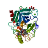 3pm3C 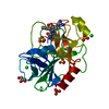 3pmjC 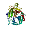 3pwbC 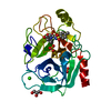 3pwcC 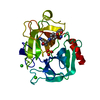 3pyhC 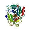 3q00C 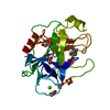 3unqC 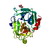 3unsC 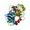 3uopC 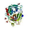 3upeC 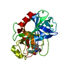 3uqoC 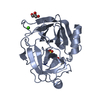 3uuzC 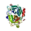 3uwiC 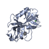 3uy9C 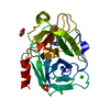 3v12C 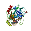 3v13C 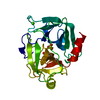 1v2kS S: Starting model for refinement C: citing same article ( |
|---|---|
| Similar structure data |


-Links
 PDBj
PDBj

- Assembly
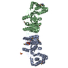
| Deposited unit | 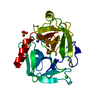
| ||||||||
|---|---|---|---|---|---|---|---|---|---|
| 1 |
| ||||||||
| Unit cell |
|
-Components
| #1: Protein | Mass: 23426.379 Da / Num. of mol.: 1 Mutation: N102E, L104Y, Y175S, P176S, G177F, Q178Y, S195A, V228F Source method: isolated from a genetically manipulated source Source: (gene. exp.)  | ||||
|---|---|---|---|---|---|
| #2: Chemical | ChemComp-CA /   Mass: 40.078 Da / Num. of mol.: 1 / Source method: obtained synthetically / Formula: Ca Mass: 40.078 Da / Num. of mol.: 1 / Source method: obtained synthetically / Formula: Ca | ||||
| #3: Chemical | ChemComp-3YH / 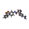  Mass: 461.493 Da / Num. of mol.: 1 / Source method: obtained synthetically / Formula: C23H19N5O4S Mass: 461.493 Da / Num. of mol.: 1 / Source method: obtained synthetically / Formula: C23H19N5O4S | ||||
| #4: Chemical |   Mass: 96.063 Da / Num. of mol.: 3 / Source method: obtained synthetically / Formula: SO4 Mass: 96.063 Da / Num. of mol.: 3 / Source method: obtained synthetically / Formula: SO4#5: Water | ChemComp-HOH / |  Mass: 18.015 Da / Num. of mol.: 171 / Source method: isolated from a natural source / Formula: H2O Mass: 18.015 Da / Num. of mol.: 171 / Source method: isolated from a natural source / Formula: H2OHas protein modification | Y | |
-Experimental details
-Experiment
| Experiment | Method: X-RAY DIFFRACTION / Number of used crystals: 1 |
|---|
- Sample preparation
| Crystal | Density Matthews: 2.26 Å3/Da / Density % sol: 45.64 % |
|---|---|
| Crystal grow | Temperature: 291 K / Method: vapor diffusion, hanging drop / pH: 7 Details: 30% PEG8000, 0.2M AMMONIUM SULPHATE, 0.1M IMIDAZOLE, pH 7.0, VAPOR DIFFUSION, HANGING DROP, temperature 291K |
-Data collection
| Diffraction | Mean temperature: 100 K |
|---|---|
| Diffraction source | Source: ROTATING ANODE / Type: RIGAKU / Wavelength: 1.5418 Å |
| Detector | Type: RIGAKU SATURN 944+ / Detector: CCD |
| Radiation | Protocol: SINGLE WAVELENGTH / Monochromatic (M) / Laue (L): M / Scattering type: x-ray |
| Radiation wavelength | Wavelength: 1.5418 Å / Relative weight: 1 |
| Reflection | Resolution: 2.4→30 Å / Num. all: 8312 / Num. obs: 8312 / % possible obs: 100 % / Observed criterion σ(F): 0 / Observed criterion σ(I): -3.7 / Redundancy: 7.6 % / Rmerge(I) obs: 0.167 / Net I/σ(I): 8.7 |
| Reflection shell | Resolution: 2.4→2.49 Å / Redundancy: 7.5 % / Rmerge(I) obs: 0.388 / Mean I/σ(I) obs: 4.5 / % possible all: 100 |
- Processing
| Software |
| ||||||||||||||||||||||||||||||||||||||||||||||||||||||||||||||||||||||||||||||||||||||||||||||||||||||||||||||||||||||||||||||||||||||||||||||||||||||||||||||||||||||||||
|---|---|---|---|---|---|---|---|---|---|---|---|---|---|---|---|---|---|---|---|---|---|---|---|---|---|---|---|---|---|---|---|---|---|---|---|---|---|---|---|---|---|---|---|---|---|---|---|---|---|---|---|---|---|---|---|---|---|---|---|---|---|---|---|---|---|---|---|---|---|---|---|---|---|---|---|---|---|---|---|---|---|---|---|---|---|---|---|---|---|---|---|---|---|---|---|---|---|---|---|---|---|---|---|---|---|---|---|---|---|---|---|---|---|---|---|---|---|---|---|---|---|---|---|---|---|---|---|---|---|---|---|---|---|---|---|---|---|---|---|---|---|---|---|---|---|---|---|---|---|---|---|---|---|---|---|---|---|---|---|---|---|---|---|---|---|---|---|---|---|---|---|
| Refinement | Method to determine structure: MOLECULAR REPLACEMENT Starting model: PDB ENTRY 1V2K Resolution: 2.4→29.81 Å / Cor.coef. Fo:Fc: 0.936 / Isotropic thermal model: Isotropic / Cross valid method: THROUGHOUT / ESU R: 0.333 / Stereochemistry target values: MAXIMUM LIKELIHOOD / Details: HYDROGENS HAVE BEEN ADDED IN THE RIDING POSITIONS
| ||||||||||||||||||||||||||||||||||||||||||||||||||||||||||||||||||||||||||||||||||||||||||||||||||||||||||||||||||||||||||||||||||||||||||||||||||||||||||||||||||||||||||
| Solvent computation | Ion probe radii: 0.8 Å / Shrinkage radii: 0.8 Å / VDW probe radii: 1.4 Å / Solvent model: MASK | ||||||||||||||||||||||||||||||||||||||||||||||||||||||||||||||||||||||||||||||||||||||||||||||||||||||||||||||||||||||||||||||||||||||||||||||||||||||||||||||||||||||||||
| Displacement parameters | Biso mean: 21.16 Å2
| ||||||||||||||||||||||||||||||||||||||||||||||||||||||||||||||||||||||||||||||||||||||||||||||||||||||||||||||||||||||||||||||||||||||||||||||||||||||||||||||||||||||||||
| Refinement step | Cycle: LAST / Resolution: 2.4→29.81 Å
| ||||||||||||||||||||||||||||||||||||||||||||||||||||||||||||||||||||||||||||||||||||||||||||||||||||||||||||||||||||||||||||||||||||||||||||||||||||||||||||||||||||||||||
| Refine LS restraints |
|
