 Movie
Movie Controller


+ Open data

- Basic information
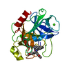
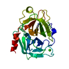
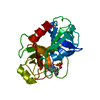
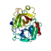
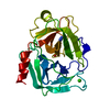
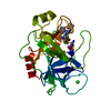
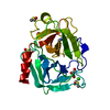
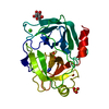
| Entry | Database: PDB / ID: 1y3x | ||||||
|---|---|---|---|---|---|---|---|
| Title | TRYPSIN INHIBITOR COMPLEX | ||||||
 Components Components | Trypsinogen, cationic | ||||||
 Keywords Keywords | HYDROLASE / Trypsin / Inhibitor Complex / BARREL / 6 STRANDED BETA-SHEET / BETA HAIRPIN / GREEK KEY MOTIVE | ||||||
| Function / homology |  Function and homology information Function and homology informationtrypsin / serpin family protein binding / serine protease inhibitor complex / digestion / endopeptidase activity / serine-type endopeptidase activity / proteolysis / : / metal ion binding Similarity search - Function | ||||||
| Biological species |  | ||||||
| Method |  X-RAY DIFFRACTION / MOLECULAR REPLACEMENT / Resolution: 1.7 Å X-RAY DIFFRACTION / MOLECULAR REPLACEMENT / Resolution: 1.7 Å | ||||||
 Authors Authors | Fokkens, J. / Klebe, G. | ||||||
 Citation Citation | Journal: Angew.Chem.Int.Ed.Engl. / Year: 2006 Title: A simple protocol to estimate differences in protein binding affinity for enantiomers without prior resolution of racemates. Authors: Fokkens, J. / Klebe, G. | ||||||
| History |
|
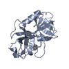
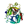
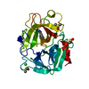
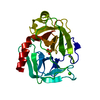
- Structure visualization
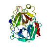
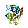
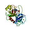
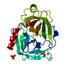
| Structure viewer | Molecule: MolmilJmol/JSmol |
|---|
- Downloads & links
-Download
| PDBx/mmCIF format | 1y3x.cif.gz | 61.3 KB | Display | PDBx/mmCIF format |
|---|---|---|---|---|
| PDB format | pdb1y3x.ent.gz | 42.4 KB | Display | PDB format |
| PDBx/mmJSON format | 1y3x.json.gz | Tree view | PDBx/mmJSON format | |
| Others |  Other downloads Other downloads |
-Validation report
| Arichive directory | https://data.pdbj.org/pub/pdb/validation_reports/y3/1y3xftp://data.pdbj.org/pub/pdb/validation_reports/y3/1y3x | HTTPS FTP |
|---|
-Related structure data
| Related structure data |  1y3vC 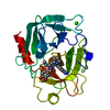 1y3wC 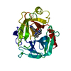 1y3yC 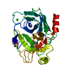 1yp9C 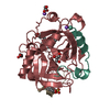 1ypeC 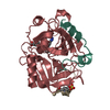 1ypgC 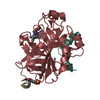 1ypjC 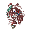 1ypkC 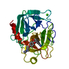 1mtsS C: citing same article ( S: Starting model for refinement |
|---|---|
| Similar structure data |


-Links
 PDBj
PDBj

- Assembly
| Deposited unit | 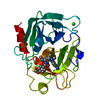
| ||||||||
|---|---|---|---|---|---|---|---|---|---|
| 1 |
| ||||||||
| Unit cell |
|
-Components
| #1: Protein | Mass: 23324.287 Da / Num. of mol.: 1 / Source method: isolated from a natural source / Source: (natural) |
|---|---|
| #2: Chemical | ChemComp-CA /   Mass: 40.078 Da / Num. of mol.: 1 / Source method: obtained synthetically / Formula: Ca Mass: 40.078 Da / Num. of mol.: 1 / Source method: obtained synthetically / Formula: Ca |
| #3: Chemical | ChemComp-UIB / (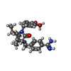  Mass: 448.557 Da / Num. of mol.: 1 / Source method: obtained synthetically / Formula: C26H32N4O3 Mass: 448.557 Da / Num. of mol.: 1 / Source method: obtained synthetically / Formula: C26H32N4O3 |
| #4: Water | ChemComp-HOH /  Mass: 18.015 Da / Num. of mol.: 150 / Source method: isolated from a natural source / Formula: H2O Mass: 18.015 Da / Num. of mol.: 150 / Source method: isolated from a natural source / Formula: H2O |
| Has protein modification | Y |
-Experimental details
-Experiment
| Experiment | Method: X-RAY DIFFRACTION / Number of used crystals: 1 |
|---|
- Sample preparation
| Crystal | Density Matthews: 2.98 Å3/Da / Density % sol: 58.73 % |
|---|---|
| Crystal grow | Temperature: 298 K / Method: vapor diffusion, hanging drop / pH: 6 Details: ammonium sulfate, MES, calcium chloride, benzamidinium hydrochloride, pH 6.00, VAPOR DIFFUSION, HANGING DROP, temperature 298K |
-Data collection
| Diffraction | Mean temperature: 293 K |
|---|---|
| Diffraction source | Source: ROTATING ANODE / Type: RIGAKU / Wavelength: 1.5418 / Wavelength: 1.5418 Å |
| Detector | Type: RIGAKU RAXIS IV / Detector: IMAGE PLATE / Date: Jul 9, 2002 / Details: OSMICS MIRRORS |
| Radiation | Monochromator: none / Protocol: SINGLE WAVELENGTH / Monochromatic (M) / Laue (L): M / Scattering type: x-ray |
| Radiation wavelength | Wavelength: 1.5418 Å / Relative weight: 1 |
| Reflection | Resolution: 1.7→47 Å / Num. all: 31574 / Num. obs: 31574 / % possible obs: 93.6 % / Observed criterion σ(F): 0 / Observed criterion σ(I): 0 / Redundancy: 2.72 % / Rmerge(I) obs: 0.058 / Rsym value: 0.058 / Net I/σ(I): 7.4 |
| Reflection shell | Resolution: 1.7→1.76 Å / Redundancy: 1.6 % / Rmerge(I) obs: 0.229 / Mean I/σ(I) obs: 1.1 / Num. unique all: 29157 / Rsym value: 0.229 / % possible all: 63.7 |
- Processing
| Software |
| |||||||||||||||||||||||||||||||||
|---|---|---|---|---|---|---|---|---|---|---|---|---|---|---|---|---|---|---|---|---|---|---|---|---|---|---|---|---|---|---|---|---|---|---|
| Refinement | Method to determine structure: MOLECULAR REPLACEMENT Starting model: PDB ENTRY 1MTS Resolution: 1.7→8 Å / Num. parameters: 7445 / Num. restraintsaints: 7103 / Cross valid method: FREE R / σ(F): 0 / σ(I): 0 / Stereochemistry target values: ENGH AND HUBER Details: ANISOTROPIC SCALING APPLIED BY THE METHOD OF PARKIN, MOEZZI & HOPE, J.APPL.CRYST.28(1995)53-56
| |||||||||||||||||||||||||||||||||
| Refine analyze | Num. disordered residues: 14 / Occupancy sum hydrogen: 1581 / Occupancy sum non hydrogen: 1806 | |||||||||||||||||||||||||||||||||
| Refinement step | Cycle: LAST / Resolution: 1.7→8 Å
| |||||||||||||||||||||||||||||||||
| Refine LS restraints |
|
