 Movie
Movie Controller

[English] 日本語
 Yorodumi
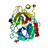
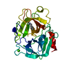
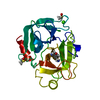
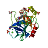
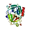
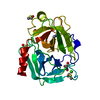
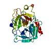
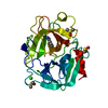
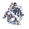
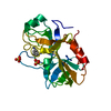
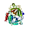
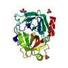
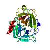
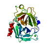
- PDB-1tgb: CRYSTAL STRUCTURE OF BOVINE TRYPSINOGEN AT 1.8 ANGSTROMS RESOLUTI... -
+ Open data

- Basic information
| Entry | Database: PDB / ID: 1tgb | ||||||
|---|---|---|---|---|---|---|---|
| Title | CRYSTAL STRUCTURE OF BOVINE TRYPSINOGEN AT 1.8 ANGSTROMS RESOLUTION. II. CRYSTALLOGRAPHIC REFINEMENT, REFINED CRYSTAL STRUCTURE AND COMPARISON WITH BOVINE TRYPSIN | ||||||
 Components Components | TRYPSINOGEN | ||||||
 Keywords Keywords | HYDROLASE ZYMOGEN (SERINE PROTEINASE) | ||||||
| Function / homology |  Function and homology information Function and homology informationtrypsin / serpin family protein binding / serine protease inhibitor complex / digestion / endopeptidase activity / serine-type endopeptidase activity / proteolysis / : / metal ion binding Similarity search - Function | ||||||
| Biological species |  | ||||||
| Method |  X-RAY DIFFRACTION / Resolution: 1.8 Å X-RAY DIFFRACTION / Resolution: 1.8 Å | ||||||
 Authors Authors | Bode, W. / Fehlhammer, H. / Huber, R. | ||||||
 Citation Citation | Journal: J.Mol.Biol. / Year: 1977 Title: Crystal structure of bovine trypsinogen at 1-8 A resolution. II. Crystallographic refinement, refined crystal structure and comparison with bovine trypsin. Authors: Fehlhammer, H. / Bode, W. / Huber, R. #1: Journal: J.Mol.Biol. / Year: 1976Title: Crystal Structure of Bovine Trypsinogen at 1.8 Angstroms Resolution. I. Data Collection, Application of Patterson Search Techniques and Preliminary Structural Interpretation Authors: Bode, W. / Fehlhammer, H. / Huber, R. #2: Journal: Acta Crystallogr.,Sect.B / Year: 1980Title: Low-Temperature Protein Crystallography. Temperature Factor, Mosaic Spread, Extinction and Diffuse Scattering in Two Examples. Bovine Trypsinogen and Fc Fragment Authors: Singh, T.P. / Bode, W. / Huber, R. #3: Journal: Acc.Chem.Res. / Year: 1978Title: Structural Basis of the Activation and Action of Trypsin Authors: Huber, R. / Bode, W. #4: Journal: FEBS Lett. / Year: 1978Title: Crystal Structure Analysis and Refinement of Two Variants of Trigonal Trypsinogen Authors: Bode, W. / Huber, R. | ||||||
| History |
|
- Structure visualization
| Structure viewer | Molecule: MolmilJmol/JSmol |
|---|
- Downloads & links
-Download
| PDBx/mmCIF format | 1tgb.cif.gz | 55.7 KB | Display | PDBx/mmCIF format |
|---|---|---|---|---|
| PDB format | pdb1tgb.ent.gz | 39.6 KB | Display | PDB format |
| PDBx/mmJSON format | 1tgb.json.gz | Tree view | PDBx/mmJSON format | |
| Others |  Other downloads Other downloads |
-Validation report
| Arichive directory | https://data.pdbj.org/pub/pdb/validation_reports/tg/1tgbftp://data.pdbj.org/pub/pdb/validation_reports/tg/1tgb | HTTPS FTP |
|---|
-Related structure data
| Similar structure data |
|---|


-Links
 PDBj
PDBj

- Assembly
| Deposited unit | 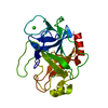
| ||||||||
|---|---|---|---|---|---|---|---|---|---|
| 1 |
| ||||||||
| Unit cell |
| ||||||||
| Atom site foot note | 1: THESE ATOMS WERE NOT FOUND IN THE ELECTRON DENSITY MAP. THEIR COORDINATES WERE GENERATED USING STEREOCHEMICAL CRITERIA. | ||||||||
| Components on special symmetry positions |
|
-Components
| #1: Protein | Mass: 24012.953 Da / Num. of mol.: 1 Source method: isolated from a genetically manipulated source Source: (gene. exp.) |
|---|---|
| #2: Chemical | ChemComp-CA /   Mass: 40.078 Da / Num. of mol.: 1 / Source method: obtained synthetically / Formula: Ca Mass: 40.078 Da / Num. of mol.: 1 / Source method: obtained synthetically / Formula: Ca |
| #3: Water | ChemComp-HOH /  Mass: 18.015 Da / Num. of mol.: 120 / Source method: isolated from a natural source / Formula: H2O Mass: 18.015 Da / Num. of mol.: 120 / Source method: isolated from a natural source / Formula: H2O |
| Has protein modification | Y |
-Experimental details
-Experiment
| Experiment | Method: X-RAY DIFFRACTION |
|---|
- Sample preparation
| Crystal | Density Matthews: 2 Å3/Da / Density % sol: 38.37 % |
|---|---|
| Crystal grow | *PLUS Method: other / Details: Bode, W., (1976) J. Mol. Biol., 106, 325. |
-Data collection
| Reflection | *PLUS Highest resolution: 1.8 Å / Num. obs: 15000 / Num. measured all: 45000 |
|---|
- Processing
| Refinement | Highest resolution: 1.8 Å Details: THE TEMPERATURE FACTOR FIELD OF THE ATOM AND HETATM RECORDS CONTAINS THE ATOMIC RADIUS WHICH HAS BEEN TENTATIVELY REFINED. THE OCCUPANCY FACTORS (WEIGHTS) HAVE BEEN REFINED ONLY FOR THE SOLVENT ATOMS. | ||||||||||||
|---|---|---|---|---|---|---|---|---|---|---|---|---|---|
| Refinement step | Cycle: LAST / Highest resolution: 1.8 Å
| ||||||||||||
| Refinement | *PLUS σ(I): 2 / Rfactor all: 0.38 / Rfactor obs: 0.27 | ||||||||||||
| Solvent computation | *PLUS | ||||||||||||
| Displacement parameters | *PLUS |
