 Movie
Movie Controller

[English] 日本語
 Yorodumi
- PDB-1tgs: THREE-DIMENSIONAL STRUCTURE OF THE COMPLEX BETWEEN PANCREATIC SEC... -
+ Open data

- Basic information
| Entry | Database: PDB / ID: 1tgs | ||||||
|---|---|---|---|---|---|---|---|
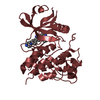
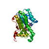
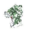
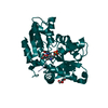
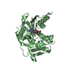
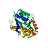
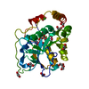
| Title | THREE-DIMENSIONAL STRUCTURE OF THE COMPLEX BETWEEN PANCREATIC SECRETORY INHIBITOR (KAZAL TYPE) AND TRYPSINOGEN AT 1.8 ANGSTROMS RESOLUTION. STRUCTURE SOLUTION, CRYSTALLOGRAPHIC REFINEMENT AND PRELIMINARY STRUCTURAL INTERPRETATION | ||||||
 Components Components |
| ||||||
 Keywords Keywords | COMPLEX (PROTEINASE/INHIBITOR) / COMPLEX (PROTEINASE-INHIBITOR) / COMPLEX (PROTEINASE-INHIBITOR) complex | ||||||
| Function / homology |  Function and homology information Function and homology informationtrypsin / serpin family protein binding / serine protease inhibitor complex / digestion / serine-type endopeptidase inhibitor activity / endopeptidase activity / serine-type endopeptidase activity / proteolysis / : / extracellular region / metal ion binding Similarity search - Function | ||||||
| Biological species |  | ||||||
| Method |  X-RAY DIFFRACTION / Resolution: 1.8 Å X-RAY DIFFRACTION / Resolution: 1.8 Å | ||||||
 Authors Authors | Bolognesi, M. / Gatti, G. / Menegatti, E. / Guarneri, M. / Marquart, M. / Papamokos, E. / Huber, R. | ||||||
 Citation Citation | Journal: J.Mol.Biol. / Year: 1982 Title: Three-dimensional structure of the complex between pancreatic secretory trypsin inhibitor (Kazal type) and trypsinogen at 1.8 A resolution. Structure solution, crystallographic refinement and ...Title: Three-dimensional structure of the complex between pancreatic secretory trypsin inhibitor (Kazal type) and trypsinogen at 1.8 A resolution. Structure solution, crystallographic refinement and preliminary structural interpretation. Authors: Bolognesi, M. / Gatti, G. / Menagatti, E. / Guarneri, M. / Marquart, M. / Papamokos, E. / Huber, R. #1: Journal: Acta Crystallogr.,Sect.B / Year: 1983Title: The Geometry of the Reactive Site and of the Peptide Groups in Trypsin, Trypsinogen and its Complexes with Inhibitors Authors: Marquart, M. / Walter, J. / Deisenhofer, J. / Bode, W. / Huber, R. | ||||||
| History |
|
- Structure visualization
| Structure viewer | Molecule: MolmilJmol/JSmol |
|---|
- Downloads & links
-Download
| PDBx/mmCIF format | 1tgs.cif.gz | 64.9 KB | Display | PDBx/mmCIF format |
|---|---|---|---|---|
| PDB format | pdb1tgs.ent.gz | 47.6 KB | Display | PDB format |
| PDBx/mmJSON format | 1tgs.json.gz | Tree view | PDBx/mmJSON format | |
| Others |  Other downloads Other downloads |
-Validation report
| Arichive directory | https://data.pdbj.org/pub/pdb/validation_reports/tg/1tgsftp://data.pdbj.org/pub/pdb/validation_reports/tg/1tgs | HTTPS FTP |
|---|
-Related structure data
| Similar structure data |
|---|


-Links
 PDBj
PDBj

- Assembly
| Deposited unit | 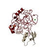
| ||||||||
|---|---|---|---|---|---|---|---|---|---|
| 1 |
| ||||||||
| Unit cell |
| ||||||||
| Atom site foot note | 1: SEE REMARK 4. |
-Components
| #1: Protein | Mass: 24012.953 Da / Num. of mol.: 1 Source method: isolated from a genetically manipulated source Source: (gene. exp.) |
|---|---|
| #2: Protein | Mass: 6028.824 Da / Num. of mol.: 1 Source method: isolated from a genetically manipulated source Source: (gene. exp.) |
| #3: Chemical | ChemComp-SO4 /   Mass: 96.063 Da / Num. of mol.: 1 / Source method: obtained synthetically / Formula: SO4 Mass: 96.063 Da / Num. of mol.: 1 / Source method: obtained synthetically / Formula: SO4 |
| #4: Chemical | ChemComp-CA /   Mass: 40.078 Da / Num. of mol.: 1 / Source method: obtained synthetically / Formula: Ca Mass: 40.078 Da / Num. of mol.: 1 / Source method: obtained synthetically / Formula: Ca |
| #5: Water | ChemComp-HOH /  Mass: 18.015 Da / Num. of mol.: 162 / Source method: isolated from a natural source / Formula: H2O Mass: 18.015 Da / Num. of mol.: 162 / Source method: isolated from a natural source / Formula: H2O |
| Has protein modification | Y |
| Sequence details | THE 229 AMINO ACIDS OF TRYPSINOGEN ARE IDENTIFIED BY THE RESIDUE NUMBERS OF THE HOMOLOGOUS ...THE 229 AMINO ACIDS OF TRYPSINOGE |
-Experimental details
-Experiment
| Experiment | Method: X-RAY DIFFRACTION |
|---|
- Sample preparation
| Crystal | Density Matthews: 2.82 Å3/Da / Density % sol: 56.37 % |
|---|---|
| Crystal grow | *PLUS Method: other / Details: Bolognesi, M., (1981) J. Mol. Biol., 145, 603. |
-Data collection
| Reflection | *PLUS Highest resolution: 1.8 Å / Num. obs: 26341 / % possible obs: 84 % / Rmerge(I) obs: 0.084 |
|---|
- Processing
| Refinement | Resolution: 1.8→7 Å / Rfactor Rwork: 0.186 | ||||||||||||
|---|---|---|---|---|---|---|---|---|---|---|---|---|---|
| Refinement step | Cycle: LAST / Resolution: 1.8→7 Å
| ||||||||||||
| Refinement | *PLUS Num. reflection obs: 26341 / Rfactor obs: 0.195 | ||||||||||||
| Solvent computation | *PLUS | ||||||||||||
| Displacement parameters | *PLUS |
