 Movie
Movie Controller


+ Open data

- Basic information
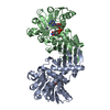
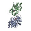
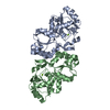
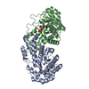
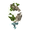
| Entry | Database: PDB / ID: 1ezx | ||||||
|---|---|---|---|---|---|---|---|
| Title | CRYSTAL STRUCTURE OF A SERPIN:PROTEASE COMPLEX | ||||||
 Components Components |
| ||||||
 Keywords Keywords | HYDROLASE/HYDROLASE INHIBITOR / protease-inhibitor complex / serpin / alpha-1-antitrypsin / trypsin / HYDROLASE-HYDROLASE INHIBITOR COMPLEX | ||||||
| Function / homology |  Function and homology information Function and homology informationCargo concentration in the ER / COPII-coated ER to Golgi transport vesicle / COPII-mediated vesicle transport / trypsin / serpin family protein binding / serine protease inhibitor complex / digestion / endoplasmic reticulum-Golgi intermediate compartment membrane / platelet alpha granule lumen / acute-phase response ...Cargo concentration in the ER / COPII-coated ER to Golgi transport vesicle / COPII-mediated vesicle transport / trypsin / serpin family protein binding / serine protease inhibitor complex / digestion / endoplasmic reticulum-Golgi intermediate compartment membrane / platelet alpha granule lumen / acute-phase response / Post-translational protein phosphorylation / serine-type endopeptidase inhibitor activity / Regulation of Insulin-like Growth Factor (IGF) transport and uptake by Insulin-like Growth Factor Binding Proteins (IGFBPs) / blood coagulation / Platelet degranulation / extracellular matrix / protease binding / endopeptidase activity / ficolin-1-rich granule lumen / endoplasmic reticulum lumen / serine-type endopeptidase activity / Neutrophil degranulation / Golgi apparatus / endoplasmic reticulum / proteolysis / : / extracellular exosome / extracellular region / metal ion binding / identical protein binding Similarity search - Function | ||||||
| Biological species |  Homo sapiens (human) Homo sapiens (human) | ||||||
| Method |  X-RAY DIFFRACTION / SYNCHROTRON / Resolution: 2.6 Å X-RAY DIFFRACTION / SYNCHROTRON / Resolution: 2.6 Å | ||||||
 Authors Authors | Huntington, J.A. / Carrell, R.W. | ||||||
 Citation Citation | Journal: Nature / Year: 2000 Title: Structure of a serpin-protease complex shows inhibition by deformation. Authors: Huntington, J.A. / Read, R.J. / Carrell, R.W. | ||||||
| History |
|
- Structure visualization
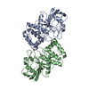
| Structure viewer | Molecule: MolmilJmol/JSmol |
|---|
- Downloads & links
-Download
| PDBx/mmCIF format | 1ezx.cif.gz | 111.9 KB | Display | PDBx/mmCIF format |
|---|---|---|---|---|
| PDB format | pdb1ezx.ent.gz | 85.4 KB | Display | PDB format |
| PDBx/mmJSON format | 1ezx.json.gz | Tree view | PDBx/mmJSON format | |
| Others |  Other downloads Other downloads |
-Validation report
| Arichive directory | https://data.pdbj.org/pub/pdb/validation_reports/ez/1ezxftp://data.pdbj.org/pub/pdb/validation_reports/ez/1ezx | HTTPS FTP |
|---|
-Related structure data
| Similar structure data |
|---|


-Links
 PDBj
PDBj


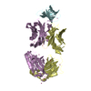
- Assembly
| Deposited unit | 
| ||||||||
|---|---|---|---|---|---|---|---|---|---|
| 1 |
| ||||||||
| Unit cell |
|
-Components
| #1: Protein | Mass: 37692.922 Da / Num. of mol.: 1 Fragment: N-TERMINAL FRAGMENT OF PROTEOLYTIC CLEAVAGE AT MET358-SER359 Source method: isolated from a genetically manipulated source Source: (gene. exp.) Homo sapiens (human) / Tissue: BLOOD / Tissue fraction: PLASMA / Plasmid: PET / Production host:  |
|---|---|
| #2: Protein/peptide | Mass: 4139.938 Da / Num. of mol.: 1 Fragment: C-TERMINAL FRAGMENT OF PROTEOLYTIC CLEAVAGE AT MET358-SER359 Source method: isolated from a genetically manipulated source Source: (gene. exp.) Homo sapiens (human) / Tissue: BLOOD / Tissue fraction: PLASMA / Plasmid: PET / Production host: |
| #3: Protein | Mass: 25444.717 Da / Num. of mol.: 1 Source method: isolated from a genetically manipulated source Source: (gene. exp.) |
| #4: Water | ChemComp-HOH /  Mass: 18.015 Da / Num. of mol.: 81 / Source method: isolated from a natural source / Formula: H2O Mass: 18.015 Da / Num. of mol.: 81 / Source method: isolated from a natural source / Formula: H2O |
| Has protein modification | Y |
-Experimental details
-Experiment
| Experiment | Method: X-RAY DIFFRACTION / Number of used crystals: 1 |
|---|
- Sample preparation
| Crystal | Density Matthews: 2.93 Å3/Da / Density % sol: 58.06 % | |||||||||||||||||||||||||
|---|---|---|---|---|---|---|---|---|---|---|---|---|---|---|---|---|---|---|---|---|---|---|---|---|---|---|
| Crystal grow | Temperature: 277 K / Method: vapor diffusion, hanging drop / pH: 8 Details: PEG 3350, sodium citrate , pH 8.0, VAPOR DIFFUSION, HANGING DROP, temperature 277K | |||||||||||||||||||||||||
| Crystal grow | *PLUS Temperature: 4 ℃ / pH: 5.7 | |||||||||||||||||||||||||
| Components of the solutions | *PLUS
|
-Data collection
| Diffraction | Mean temperature: 100 K |
|---|---|
| Diffraction source | Source: SYNCHROTRON / Site: SRS  / Beamline: PX9.6 / Wavelength: 0.87 / Beamline: PX9.6 / Wavelength: 0.87 |
| Detector | Type: ADSC QUANTUM 4 / Detector: CCD / Date: Apr 14, 2000 |
| Radiation | Protocol: SINGLE WAVELENGTH / Monochromatic (M) / Laue (L): M / Scattering type: x-ray |
| Radiation wavelength | Wavelength: 0.87 Å / Relative weight: 1 |
| Reflection | Resolution: 2.6→30 Å / Num. all: 24803 / Num. obs: 24548 / % possible obs: 99 % / Observed criterion σ(F): 2 / Observed criterion σ(I): 2 / Redundancy: 7.7 % / Biso Wilson estimate: 58.9 Å2 / Rmerge(I) obs: 0.158 / Net I/σ(I): 12 |
| Reflection shell | Resolution: 2.6→2.74 Å / Redundancy: 8 % / Rmerge(I) obs: 0.833 / Num. unique all: 3524 / % possible all: 99 |
| Reflection | *PLUS Num. measured all: 190117 |
| Reflection shell | *PLUS % possible obs: 99 % |
- Processing
| Software |
| |||||||||||||||||||||||||
|---|---|---|---|---|---|---|---|---|---|---|---|---|---|---|---|---|---|---|---|---|---|---|---|---|---|---|
| Refinement | Resolution: 2.6→30 Å / σ(F): 1 / σ(I): 1 / Stereochemistry target values: Engh & Huber / Details: maximum likelihood target of Pannu and Read, 1996
| |||||||||||||||||||||||||
| Refinement step | Cycle: LAST / Resolution: 2.6→30 Å
| |||||||||||||||||||||||||
| Refine LS restraints |
|
