 Movie
Movie Controller


+ Open data

- Basic information
| Entry | Database: PDB / ID: 1yfo | ||||||
|---|---|---|---|---|---|---|---|
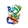
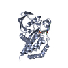
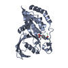
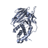
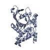
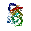
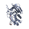
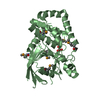
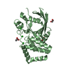
| Title | RECEPTOR PROTEIN TYROSINE PHOSPHATASE ALPHA, DOMAIN 1 FROM MOUSE | ||||||
 Components Components | RECEPTOR PROTEIN TYROSINE PHOSPHATASE ALPHA | ||||||
 Keywords Keywords | HYDROLASE / SIGNAL TRANSDUCTION / RECEPTOR / GLYCOPROTEIN / PHOSPHORYLATION | ||||||
| Function / homology |  Function and homology information Function and homology informationNCAM signaling for neurite out-growth / RAF/MAP kinase cascade / regulation of focal adhesion assembly / positive regulation of oligodendrocyte differentiation / protein-tyrosine-phosphatase / protein tyrosine phosphatase activity / integrin-mediated signaling pathway / synaptic membrane / modulation of chemical synaptic transmission / Schaffer collateral - CA1 synapse ...NCAM signaling for neurite out-growth / RAF/MAP kinase cascade / regulation of focal adhesion assembly / positive regulation of oligodendrocyte differentiation / protein-tyrosine-phosphatase / protein tyrosine phosphatase activity / integrin-mediated signaling pathway / synaptic membrane / modulation of chemical synaptic transmission / Schaffer collateral - CA1 synapse / insulin receptor signaling pathway / protein phosphorylation / signaling receptor complex / focal adhesion / protein-containing complex binding / signal transduction / membrane / identical protein binding Similarity search - Function | ||||||
| Biological species |  | ||||||
| Method |  X-RAY DIFFRACTION / MOLECULAR REPLACEMENT / Resolution: 2.25 Å X-RAY DIFFRACTION / MOLECULAR REPLACEMENT / Resolution: 2.25 Å | ||||||
 Authors Authors | Bilwes, A.M. / Noel, J.P. | ||||||
 Citation Citation | Journal: Nature / Year: 1996 Title: Structural basis for inhibition of receptor protein-tyrosine phosphatase-alpha by dimerization. Authors: Bilwes, A.M. / den Hertog, J. / Hunter, T. / Noel, J.P. | ||||||
| History |
|
- Structure visualization
| Structure viewer | Molecule: MolmilJmol/JSmol |
|---|
- Downloads & links
-Download
| PDBx/mmCIF format | 1yfo.cif.gz | 130.4 KB | Display | PDBx/mmCIF format |
|---|---|---|---|---|
| PDB format | pdb1yfo.ent.gz | 101.1 KB | Display | PDB format |
| PDBx/mmJSON format | 1yfo.json.gz | Tree view | PDBx/mmJSON format | |
| Others |  Other downloads Other downloads |
-Validation report
| Arichive directory | https://data.pdbj.org/pub/pdb/validation_reports/yf/1yfoftp://data.pdbj.org/pub/pdb/validation_reports/yf/1yfo | HTTPS FTP |
|---|
-Related structure data
| Related structure data | 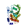 2hnpS S: Starting model for refinement |
|---|---|
| Similar structure data |

-Links
 PDBj
PDBj


- Assembly
| Deposited unit | 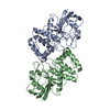
| ||||||||
|---|---|---|---|---|---|---|---|---|---|
| 1 | 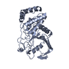
| ||||||||
| 2 | 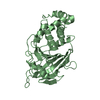
| ||||||||
| Unit cell |
| ||||||||
| Noncrystallographic symmetry (NCS) | NCS oper: (Code: given Matrix: (0.999983, 0.001238, -0.005715), Vector: |
-Components
| #1: Protein | Mass: 34894.770 Da / Num. of mol.: 2 / Fragment: MEMBRANE PROXIMAL CATALYTIC DOMAIN Source method: isolated from a genetically manipulated source Source: (gene. exp.)  #2: Water | ChemComp-HOH / |  Mass: 18.015 Da / Num. of mol.: 287 / Source method: isolated from a natural source / Formula: H2O Mass: 18.015 Da / Num. of mol.: 287 / Source method: isolated from a natural source / Formula: H2OCompound details | RESIDUES 379 - 386 WERE NOT MODELLED FOR EACH MONOMER TRYPSIN CUT AT 385 OR 386. | |
|---|
-Experimental details
-Experiment
| Experiment | Method: X-RAY DIFFRACTION / Number of used crystals: 1 |
|---|
- Sample preparation
| Crystal | Density Matthews: 2.08 Å3/Da / Density % sol: 44 % | |||||||||||||||||||||||||
|---|---|---|---|---|---|---|---|---|---|---|---|---|---|---|---|---|---|---|---|---|---|---|---|---|---|---|
| Crystal grow | pH: 5 Details: PROTEIN WAS CRYSTALLIZED FROM 12% PEG 8000 0.3 M AMMONIUM ACETATE, 50 MM SODIUM ACETATE PH 5, 2 MM DTT. ETHYLENE GLYCOL 20% WAS USED AS A CRYOPROTECTANT., pH 5.0 | |||||||||||||||||||||||||
| Crystal grow | *PLUS Method: vapor diffusion, hanging drop | |||||||||||||||||||||||||
| Components of the solutions | *PLUS
|
-Data collection
| Diffraction | Mean temperature: 100 K |
|---|---|
| Diffraction source | Source: ROTATING ANODE / Type: MACSCIENCE M18X / Wavelength: 1.5418 |
| Detector | Type: MACSCIENCE / Detector: IMAGE PLATE / Date: Jul 14, 1995 / Details: MIRRORS |
| Radiation | Monochromator: NI FILTER / Monochromatic (M) / Laue (L): M / Scattering type: x-ray |
| Radiation wavelength | Wavelength: 1.5418 Å / Relative weight: 1 |
| Reflection | Resolution: 2.25→23 Å / Num. obs: 22317 / % possible obs: 82.4 % / Observed criterion σ(I): 0 / Redundancy: 2.6 % / Biso Wilson estimate: 27 Å2 / Rsym value: 0.046 / Net I/σ(I): 22 |
| Reflection shell | Resolution: 2.25→2.35 Å / Redundancy: 1 % / Mean I/σ(I) obs: 7 / Rsym value: 0.104 / % possible all: 17.4 |
| Reflection | *PLUS Rmerge(I) obs: 0.046 |
| Reflection shell | *PLUS % possible obs: 17.4 % / Rmerge(I) obs: 0.104 |
- Processing
| Software |
| ||||||||||||||||||||||||||||||||||||||||||||||||||||||||||||
|---|---|---|---|---|---|---|---|---|---|---|---|---|---|---|---|---|---|---|---|---|---|---|---|---|---|---|---|---|---|---|---|---|---|---|---|---|---|---|---|---|---|---|---|---|---|---|---|---|---|---|---|---|---|---|---|---|---|---|---|---|---|
| Refinement | Method to determine structure: MOLECULAR REPLACEMENT Starting model: PDB ENTRY 2HNP Resolution: 2.25→20 Å / σ(F): 0
| ||||||||||||||||||||||||||||||||||||||||||||||||||||||||||||
| Refinement step | Cycle: LAST / Resolution: 2.25→20 Å
| ||||||||||||||||||||||||||||||||||||||||||||||||||||||||||||
| Refine LS restraints |
| ||||||||||||||||||||||||||||||||||||||||||||||||||||||||||||
| LS refinement shell | Resolution: 2.25→2.35 Å / Total num. of bins used: 8
| ||||||||||||||||||||||||||||||||||||||||||||||||||||||||||||
| Xplor file |
| ||||||||||||||||||||||||||||||||||||||||||||||||||||||||||||
| Software | *PLUS Name: X-PLOR / Classification: refinement | ||||||||||||||||||||||||||||||||||||||||||||||||||||||||||||
| Refinement | *PLUS | ||||||||||||||||||||||||||||||||||||||||||||||||||||||||||||
| Solvent computation | *PLUS | ||||||||||||||||||||||||||||||||||||||||||||||||||||||||||||
| Displacement parameters | *PLUS | ||||||||||||||||||||||||||||||||||||||||||||||||||||||||||||
| Refine LS restraints | *PLUS
| ||||||||||||||||||||||||||||||||||||||||||||||||||||||||||||
| LS refinement shell | *PLUS Rfactor obs: 0.281 |
