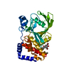
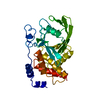
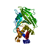
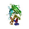
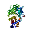
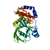
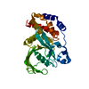
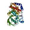
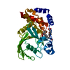
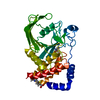
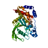
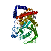
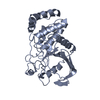
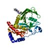
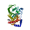
Entry Database : PDB / ID : 1t49Title Allosteric Inhibition of Protein Tyrosine Phosphatase 1B Protein-tyrosine phosphatase, non-receptor type 1 Keywords / / Function / homology Function Domain/homology Component
/ / / / / / / / / / / / / / / / / / / / / / / / / / / / / / / / / / / / / / / / / / / / / / / / / / / / / / / / / / / / / / / / / / / / / / / / / / / / / / / / / / / / / / / / / / / / / / / / / / / / / / / / / / / / / / Biological species Homo sapiens (human)Method / / Resolution : 1.9 Å Authors Wiesmann, C. / Barr, K.J. / Kung, J. / Zhu, J. / Shen, W. / Fahr, B.J. / Zhong, M. / Taylor, L. / Randal, M. / McDowell, R.S. / Hansen, S.K. Journal : Nat.Struct.Mol.Biol. / Year : 2004Title : Allosteric inhibition of protein tyrosine phosphatase 1B.Authors : Wiesmann, C. / Barr, K.J. / Kung, J. / Zhu, J. / Erlanson, D.A. / Shen, W. / Fahr, B.J. / Zhong, M. / Taylor, L. / Randal, M. / McDowell, R.S. / Hansen, S.K. History Deposition Apr 28, 2004 Deposition site / Processing site Revision 1.0 Jul 20, 2004 Provider / Type Revision 1.1 Apr 30, 2008 Group Revision 1.2 Jul 13, 2011 Group Revision 1.3 Feb 14, 2024 Group / Database references / Derived calculationsCategory chem_comp_atom / chem_comp_bond ... chem_comp_atom / chem_comp_bond / database_2 / pdbx_struct_conn_angle / struct_conn / struct_site Item _database_2.pdbx_DOI / _database_2.pdbx_database_accession ... _database_2.pdbx_DOI / _database_2.pdbx_database_accession / _pdbx_struct_conn_angle.ptnr1_auth_seq_id / _pdbx_struct_conn_angle.ptnr1_symmetry / _pdbx_struct_conn_angle.ptnr3_auth_seq_id / _pdbx_struct_conn_angle.ptnr3_symmetry / _pdbx_struct_conn_angle.value / _struct_conn.pdbx_dist_value / _struct_conn.ptnr2_auth_seq_id / _struct_conn.ptnr2_symmetry / _struct_site.pdbx_auth_asym_id / _struct_site.pdbx_auth_comp_id / _struct_site.pdbx_auth_seq_id
Show all Show less
 Movie
Movie Controller


 Open data
 Basic information
 Components
 Keywords
 Function and homology information
 Homo sapiens (human)
 X-RAY DIFFRACTION /
  Authors
 Citation
 Structure visualization
 Downloads & links
 Other downloads
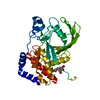


 PDBj
PDBj


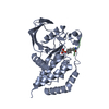
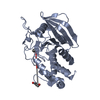
 Assembly
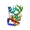


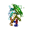
 Mass: 24.305 Da / Num. of mol.: 1 / Source method: obtained synthetically / Formula: Mg
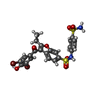
 Mass: 658.336 Da / Num. of mol.: 1 / Source method: obtained synthetically / Formula: C23H18Br2N2O7S2
 Mass: 18.015 Da / Num. of mol.: 235 / Source method: isolated from a natural source / Formula: H2O
 Sample preparation
 Processing