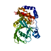
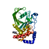
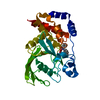
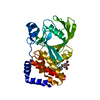
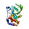
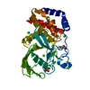
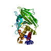
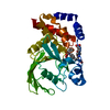
Entry Database : PDB / ID : 6cwvTitle Protein Tyrosine Phosphatase 1B A122S mutant Tyrosine-protein phosphatase non-receptor type 1 Keywords / / / / Function / homology Function Domain/homology Component
/ / / / / / / / / / / / / / / / / / / / / / / / / / / / / / / / / / / / / / / / / / / / / / / / / / / / / / / / / / / / / / / / / / / / / / / / / / / / / / / / / / / / / / / / / / / / / / / / / / / / / / / / / / / / / / Biological species Homo sapiens (human)Method / / / Resolution : 1.98002287694 Å Authors Hjortness, M. / Zwart, P. / Sankaran, B. / Fox, J.M. Funding support Organization Grant number Country National Science Foundation (NSF, United States) 1750244 National Science Foundation (NSF, United States) 1804897
Journal : Biochemistry / Year : 2018Title : Evolutionarily Conserved Allosteric Communication in Protein Tyrosine Phosphatases.Authors : Hjortness, M.K. / Riccardi, L. / Hongdusit, A. / Zwart, P.H. / Sankaran, B. / De Vivo, M. / Fox, J.M. History Deposition Mar 31, 2018 Deposition site / Processing site Revision 1.0 Oct 24, 2018 Provider / Type Revision 1.1 Oct 31, 2018 Group / Database references / Category / citation_authorItem _citation.journal_id_CSD / _citation.journal_id_ISSN ... _citation.journal_id_CSD / _citation.journal_id_ISSN / _citation.pdbx_database_id_DOI / _citation.pdbx_database_id_PubMed / _citation.title / _citation_author.name Revision 1.2 Nov 21, 2018 Group / Database references / Category / citation_authorItem _citation.journal_volume / _citation.page_first ... _citation.journal_volume / _citation.page_first / _citation.page_last / _citation_author.identifier_ORCID Revision 1.3 Nov 27, 2019 Group / Category / Item Revision 1.4 Oct 4, 2023 Group / Database references / Refinement descriptionCategory chem_comp_atom / chem_comp_bond ... chem_comp_atom / chem_comp_bond / database_2 / pdbx_initial_refinement_model Item / _database_2.pdbx_database_accession
Show all Show less
 Movie
Movie Controller


 Open data
 Basic information
 Components
 Keywords
 Function and homology information
 Homo sapiens (human)
 X-RAY DIFFRACTION /
  Authors
 United States, 2items
  Citation
 Structure visualization
 Downloads & links
 Other downloads
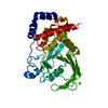


 PDBj
PDBj


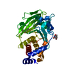
 Assembly
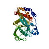


 Mass: 24.305 Da / Num. of mol.: 1 / Source method: obtained synthetically / Formula: Mg
 Mass: 18.015 Da / Num. of mol.: 220 / Source method: isolated from a natural source / Formula: H2O
 Sample preparation
 Processing