[English] 日本語
Yorodumi
- PDB-1j8a: CRYSTAL STRUCTURE OF BENZAMIDINE INHIBITED BOVINE PANCREATIC TRYP... -

+
Open data


ID or keywords:

Loading...

-
Basic information

Entry
Database: PDB / ID: 1j8a
Title
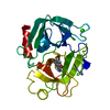
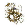
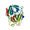
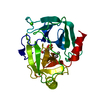
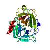
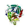
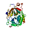
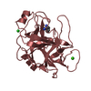
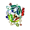
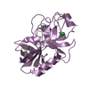
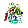
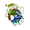
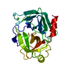
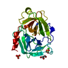
CRYSTAL STRUCTURE OF BENZAMIDINE INHIBITED BOVINE PANCREATIC TRYPSIN AT 105K TO 1.21A RESOLUTION FROM LABORATORY SOURCE WITH HIGH NUMBER OF WATERS MODELLED
ComponentsTRYPSINOGEN, CATIONIC
KeywordsHYDROLASE / PROTEIN-INHIBITOR COMPLEX / HIGH NUMBER OF WATERS / ATOMIC RESOLUTION
Function / homology
Function and homology information


trypsin / serpin family protein binding / serine protease inhibitor complex / digestion / endopeptidase activity / serine-type endopeptidase activity / proteolysis / extracellular space / metal ion binding
Similarity search - Function
Serine proteases, trypsin family, histidine active site / Serine proteases, trypsin family, serine active site / Peptidase S1A, chymotrypsin family / Serine proteases, trypsin family, histidine active site. / Serine proteases, trypsin domain profile. / Serine proteases, trypsin family, serine active site. / Trypsin-like serine protease / Serine proteases, trypsin domain / Trypsin / Trypsin-like serine proteases ...Serine proteases, trypsin family, histidine active site / Serine proteases, trypsin family, serine active site / Peptidase S1A, chymotrypsin family / Serine proteases, trypsin family, histidine active site. / Serine proteases, trypsin domain profile. / Serine proteases, trypsin family, serine active site. / Trypsin-like serine protease / Serine proteases, trypsin domain / Trypsin / Trypsin-like serine proteases / Thrombin, subunit H / Peptidase S1, PA clan, chymotrypsin-like fold / Peptidase S1, PA clan / Beta Barrel / Mainly Beta
Similarity search - Domain/homology
BENZAMIDINE / Serine protease 1
Similarity search - Component
Biological speciesBos taurus (cattle)
MethodX-RAY DIFFRACTION / MOLECULAR REPLACEMENT / Resolution: 1.21 Å
AuthorsCuesta-Seijo, J.A. / Garcia-Granda, S.
CitationJournal: BOL.R.SOC.HIST.NAT.SEC.GEOL. / Year: 2002
Title: Trypsin as a model for high resolution X-ray diffraction in proteins.
Authors: Cuesta-Seijo, J.A. / Garcia-Granda, S.
History
DepositionMay 21, 2001Deposition site: RCSB / Processing site: RCSB
Revision 1.0Sep 12, 2001Provider: repository / Type: Initial release
Revision 1.1Apr 27, 2008Group: Version format compliance
Revision 1.2Jul 13, 2011Group: Non-polymer description / Version format compliance
Revision 1.3Aug 16, 2023Group: Data collection / Database references ...Data collection / Database references / Derived calculations / Refinement description
Category: chem_comp_atom / chem_comp_bond ...chem_comp_atom / chem_comp_bond / database_2 / pdbx_initial_refinement_model / pdbx_struct_conn_angle / struct_conn / struct_site
Item: _database_2.pdbx_DOI / _database_2.pdbx_database_accession ..._database_2.pdbx_DOI / _database_2.pdbx_database_accession / _pdbx_struct_conn_angle.ptnr1_auth_comp_id / _pdbx_struct_conn_angle.ptnr1_auth_seq_id / _pdbx_struct_conn_angle.ptnr1_label_asym_id / _pdbx_struct_conn_angle.ptnr1_label_atom_id / _pdbx_struct_conn_angle.ptnr1_label_comp_id / _pdbx_struct_conn_angle.ptnr1_label_seq_id / _pdbx_struct_conn_angle.ptnr3_auth_comp_id / _pdbx_struct_conn_angle.ptnr3_auth_seq_id / _pdbx_struct_conn_angle.ptnr3_label_asym_id / _pdbx_struct_conn_angle.ptnr3_label_atom_id / _pdbx_struct_conn_angle.ptnr3_label_comp_id / _pdbx_struct_conn_angle.ptnr3_label_seq_id / _pdbx_struct_conn_angle.value / _struct_conn.pdbx_dist_value / _struct_conn.ptnr1_auth_comp_id / _struct_conn.ptnr1_auth_seq_id / _struct_conn.ptnr1_label_asym_id / _struct_conn.ptnr1_label_atom_id / _struct_conn.ptnr1_label_comp_id / _struct_conn.ptnr1_label_seq_id / _struct_conn.ptnr2_auth_comp_id / _struct_conn.ptnr2_auth_seq_id / _struct_conn.ptnr2_label_asym_id / _struct_conn.ptnr2_label_atom_id / _struct_conn.ptnr2_label_comp_id / _struct_conn.ptnr2_label_seq_id / _struct_site.pdbx_auth_asym_id / _struct_site.pdbx_auth_comp_id / _struct_site.pdbx_auth_seq_id

-
Structure visualization

Structure viewerMolecule:
MolmilJmol/JSmol

Downloads & links

-
Assembly

Deposited unit
A: TRYPSINOGEN, CATIONIC
hetero molecules


Theoretical massNumber of molelcules
Total (without water)24,0799
Polymers23,3241
Non-polymers7558
Water6,936385
1


  • Idetical with deposited unit
  • defined by author
TypeNameSymmetry operationNumber
identity operation1_555x,y,z1
Unit cell
Length a, b, c (Å)54.275, 58.568, 66.141
Angle α, β, γ (deg.)90.00, 90.00, 90.00
Int Tables number19
Space group name H-MP212121

-
Components

-
Protein , 1 types, 1 molecules A

#1: Protein TRYPSINOGEN, CATIONIC / / BETA-TRYPSIN


Mass: 23324.287 Da / Num. of mol.: 1 / Source method: isolated from a natural source / Details: bovine pancreas / Source: (natural) Bos taurus (cattle) / References: UniProt: P00760, trypsin

-
Non-polymers , 6 types, 393 molecules

#2: Chemical ChemComp-CA / CALCIUM ION


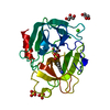
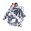
Mass: 40.078 Da / Num. of mol.: 1 / Source method: obtained synthetically / Formula: Ca
#3: Chemical ChemComp-SO4 / SULFATE ION / Sulfate


Mass: 96.063 Da / Num. of mol.: 3 / Source method: obtained synthetically / Formula: SO4
#4: Chemical ChemComp-BEN / BENZAMIDINE / Benzamidine


Mass: 120.152 Da / Num. of mol.: 1 / Source method: obtained synthetically / Formula: C7H8N2
#5: Chemical ChemComp-TRS / 2-AMINO-2-HYDROXYMETHYL-PROPANE-1,3-DIOL / TRIS BUFFER / Tris


Mass: 122.143 Da / Num. of mol.: 1 / Source method: obtained synthetically / Formula: C4H12NO3 / Comment: pH buffer*YM
#6: Chemical ChemComp-GOL / GLYCEROL / GLYCERIN / PROPANE-1,2,3-TRIOL / Glycerol


Mass: 92.094 Da / Num. of mol.: 2 / Source method: obtained synthetically / Formula: C3H8O3
#7: Water ChemComp-HOH / water / Water


Mass: 18.015 Da / Num. of mol.: 385 / Source method: isolated from a natural source / Formula: H2O

-
Experimental details

-
Experiment

ExperimentMethod: X-RAY DIFFRACTION / Number of used crystals: 1

-
Sample preparation

CrystalDensity Matthews: 2.25 Å3/Da / Density % sol: 45.42 %
Crystal growTemperature: 293.15 K / Method: vapor diffusion, hanging drop / pH: 7.4
Details: PEG 6000, ammonium sulfate, tris-HCl, benzamidine, calcium chloride, pH 7.4, VAPOR DIFFUSION, HANGING DROP, temperature 293.15K

-
Data collection

DiffractionMean temperature: 105 K
Diffraction sourceSource: ROTATING ANODE / Type: ENRAF-NONIUS FR590 / Wavelength: 1.5418
DetectorType: ENRAF-NONIUS / Detector: ENRAF-NONIUS / Date: Jun 9, 2000
RadiationMonochromator: GRAPHITE / Protocol: SINGLE WAVELENGTH / Monochromatic (M) / Laue (L): M / Scattering type: x-ray
Radiation wavelengthWavelength: 1.5418 Å / Relative weight: 1
ReflectionResolution: 1.2→19.9 Å / Num. all: 64859 / Num. obs: 64859 / % possible obs: 97.4 % / Observed criterion σ(F): 0 / Observed criterion σ(I): 0 / Redundancy: 10.5 % / Biso Wilson estimate: 11.6 Å2 / Rmerge(I) obs: 0.061 / Net I/σ(I): 13.2
Reflection shellResolution: 1.2→1.24 Å / Redundancy: 2.1 % / Rmerge(I) obs: 0.429 / Mean I/σ(I) obs: 1.5 / Num. unique all: 6239 / % possible all: 77.1

-
Processing

Software
NameVersionClassification
DENZOdata reduction
SCALEPACKdata scaling
AMoREphasing
CNS1refinement
RefinementMethod to determine structure: MOLECULAR REPLACEMENT
Starting model: PDB ENTRY 5PTP
Resolution: 1.21→19.9 Å / Isotropic thermal model: Isotropic / Cross valid method: THROUGHOUT / σ(F): 0 / σ(I): 0 / Stereochemistry target values: Engh & Huber
Details: Used maximum likelihood with structure factors target from CNS
RfactorNum. reflection% reflectionSelection details
Rfree0.178 3273 -RANDOM
Rwork0.16 ---
all0.161 64546 --
obs0.161 64546 99.4 %-
Displacement parametersBiso mean: 9.5 Å2
Baniso -1Baniso -2Baniso -3
1--8.1 Å20 Å20.674 Å2
2--0 Å20.138 Å2
3---0 Å2
Refine analyze
FreeObs
Luzzati coordinate error0.13 Å0.12 Å
Luzzati d res low-5 Å
Luzzati sigma a0.13 Å0.13 Å
Refinement stepCycle: LAST / Resolution: 1.21→19.9 Å
ProteinNucleic acidLigandSolventTotal
Num. atoms1629 0 45 385 2059
Refine LS restraints
Refine-IDTypeDev ideal
X-RAY DIFFRACTIONc_angle_deg1.574
X-RAY DIFFRACTIONc_bond_d0.00839
X-RAY DIFFRACTIONc_dihedral_angle_deg25.877
X-RAY DIFFRACTIONc_improper_angle_deg1.023
X-RAY DIFFRACTIONc_mcangle_it1.077
X-RAY DIFFRACTIONc_mcbond_it0.753
X-RAY DIFFRACTIONc_scangle_it1.908
X-RAY DIFFRACTIONc_scbond_it1.453
LS refinement shell
Resolution (Å)Rfactor RfreeNum. reflection RfreeRfactor RworkRefine-IDNum. reflection obs% reflection obs (%)
1.21-1.250.2763430.253X-RAY DIFFRACTION623997.5
1.25-1.30.2543330.235X-RAY DIFFRACTION636499.1
1.3-1.360.2023220.213X-RAY DIFFRACTION639899.6
1.36-1.430.1953390.185X-RAY DIFFRACTION640699.7
1.43-1.520.1712990.161X-RAY DIFFRACTION641399.6
1.52-1.640.1563510.147X-RAY DIFFRACTION644199.5
1.64-1.810.1613050.141X-RAY DIFFRACTION643499.4
1.81-2.070.1723230.144X-RAY DIFFRACTION649799.8
2.07-2.610.1643500.14X-RAY DIFFRACTION656599.8
2.61-19.90.163080.146X-RAY DIFFRACTION678999.7

+
About Yorodumi

-
News

-
Feb 9, 2022. New format data for meta-information of EMDB entries

New format data for meta-information of EMDB entries

  • Version 3 of the EMDB header file is now the official format.
  • The previous official version 1.9 will be removed from the archive.

Related info.:EMDB header

External links:wwPDB to switch to version 3 of the EMDB data model

-
Aug 12, 2020. Covid-19 info

Covid-19 info

URL: https://pdbj.org/emnavi/covid19.php

New page: Covid-19 featured information page in EM Navigator.

Related info.:Covid-19 info / Mar 5, 2020. Novel coronavirus structure data

+
Mar 5, 2020. Novel coronavirus structure data

Novel coronavirus structure data

Related info.:Yorodumi Speices / Aug 12, 2020. Covid-19 info

External links:COVID-19 featured content - PDBj / Molecule of the Month (242):Coronavirus Proteases

+
Jan 31, 2019. EMDB accession codes are about to change! (news from PDBe EMDB page)

EMDB accession codes are about to change! (news from PDBe EMDB page)

  • The allocation of 4 digits for EMDB accession codes will soon come to an end. Whilst these codes will remain in use, new EMDB accession codes will include an additional digit and will expand incrementally as the available range of codes is exhausted. The current 4-digit format prefixed with “EMD-” (i.e. EMD-XXXX) will advance to a 5-digit format (i.e. EMD-XXXXX), and so on. It is currently estimated that the 4-digit codes will be depleted around Spring 2019, at which point the 5-digit format will come into force.
  • The EM Navigator/Yorodumi systems omit the EMD- prefix.

Related info.:Q: What is EMD? / ID/Accession-code notation in Yorodumi/EM Navigator

External links:EMDB Accession Codes are Changing Soon! / Contact to PDBj

+
Jul 12, 2017. Major update of PDB

Major update of PDB

  • wwPDB released updated PDB data conforming to the new PDBx/mmCIF dictionary.
  • This is a major update changing the version number from 4 to 5, and with Remediation, in which all the entries are updated.
  • In this update, many items about electron microscopy experimental information are reorganized (e.g. em_software).
  • Now, EM Navigator and Yorodumi are based on the updated data.

External links:wwPDB Remediation / Enriched Model Files Conforming to OneDep Data Standards Now Available in the PDB FTP Archive

-
Yorodumi

Thousand views of thousand structures

  • Yorodumi is a browser for structure data from EMDB, PDB, SASBDB, etc.
  • This page is also the successor to EM Navigator detail page, and also detail information page/front-end page for Omokage search.
  • The word "yorodu" (or yorozu) is an old Japanese word meaning "ten thousand". "mi" (miru) is to see.

Related info.:EMDB / PDB / SASBDB / Comparison of 3 databanks / Yorodumi Search / Aug 31, 2016. New EM Navigator & Yorodumi / Yorodumi Papers / Jmol/JSmol / Function and homology information / Changes in new EM Navigator and Yorodumi

Read more