 Movie
Movie Controller


+ Open data

- Basic information
| Entry | Database: PDB / ID: 5ptp | |||||||||
|---|---|---|---|---|---|---|---|---|---|---|
| Title | STRUCTURE OF HYDROLASE (SERINE PROTEINASE) | |||||||||
 Components Components | BETA TRYPSIN | |||||||||
 Keywords Keywords | SERINE PROTEASE / HYDROLASE / DIGESTION / PANCREAS / ZYMOGEN | |||||||||
| Function / homology |  Function and homology information Function and homology informationtrypsin / serpin family protein binding / serine protease inhibitor complex / digestion / endopeptidase activity / serine-type endopeptidase activity / proteolysis / : / metal ion binding Similarity search - Function | |||||||||
| Biological species |  | |||||||||
| Method |  X-RAY DIFFRACTION / Resolution: 1.34 Å X-RAY DIFFRACTION / Resolution: 1.34 Å | |||||||||
 Authors Authors | Stroud, R.M. / Finer-Moore, J. | |||||||||
 Citation Citation | Journal: Proteins / Year: 1992 Title: Solvent structure in crystals of trypsin determined by X-ray and neutron diffraction. Authors: Finer-Moore, J.S. / Kossiakoff, A.A. / Hurley, J.H. / Earnest, T. / Stroud, R.M. #1: Journal: Acta Crystallogr.,Sect.B / Year: 1979Title: The Accuracy of Refined Protein Structures, Comparison of Two Independently Refined Models of Bovine Trypsin Authors: Chambers, J.L. / Stroud, R.M. #2: Journal: Acta Crystallogr.,Sect.B / Year: 1977Title: Difference-Fourier Refinement of the Structure of Dip-Trypsin at 1.5 Angstroms Using a Minicomputer Technique Authors: Chambers, J.L. / Stroud, R.M. #3: Journal: PROTEASES AND BIOLOGICAL CONTROL / Year: 1975Title: Structure-Function Relationships in the Serine Proteases Authors: Stroud, R.M. / Krieger, M. / Koeppe II, R.E. / Kossiakoff, A.A. / Chambers, J.L. #4: Journal: Biochem.Biophys.Res.Commun. / Year: 1974Title: Silver Ion Inhibition of Serine Proteases: Crystallographic Study of Silver-Trypsin Authors: Chambers, J.L. / Christoph, G.G. / Krieger, M. / Kay, L. / Stroud, R.M. #5: Journal: J.Mol.Biol. / Year: 1974Title: The Structure of Bovine Trypsin: Electron Density Maps of the Inhibited Enzyme at 5 a and at 2.7 A Resolution Authors: Stroud, R.M. / Kay, L.M. / Dickerson, R.E. #6: Journal: J.Mol.Biol. / Year: 1974Title: Structure and Specific Binding of Trypsin: Comparison of Inhibited Derivatives and a Model for Substrate Binding Authors: Krieger, M. / Kay, L.M. / Stroud, R.M. #7: Journal: Cold Spring Harbor Symp.Quant.Biol. / Year: 1972Title: The Crystal and Molecular Structure of Dip-Inhibited Bovine Trypsin at 2.7 A Resolution Authors: Stroud, R.M. / Kay, L.M. / Dickerson, R.E. | |||||||||
| History |
|
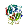
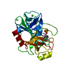
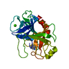
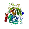
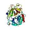
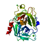
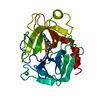
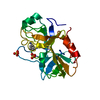
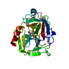
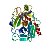
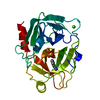
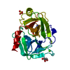
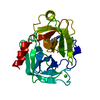
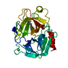
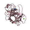
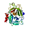
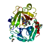
- Structure visualization
| Structure viewer | Molecule: MolmilJmol/JSmol |
|---|
- Downloads & links
-Download
| PDBx/mmCIF format | 5ptp.cif.gz | 60.6 KB | Display | PDBx/mmCIF format |
|---|---|---|---|---|
| PDB format | pdb5ptp.ent.gz | 43.7 KB | Display | PDB format |
| PDBx/mmJSON format | 5ptp.json.gz | Tree view | PDBx/mmJSON format | |
| Others |  Other downloads Other downloads |
-Validation report
| Arichive directory | https://data.pdbj.org/pub/pdb/validation_reports/pt/5ptpftp://data.pdbj.org/pub/pdb/validation_reports/pt/5ptp | HTTPS FTP |
|---|
-Related structure data
| Similar structure data |
|---|


-Links
 PDBj
PDBj

- Assembly
| Deposited unit | 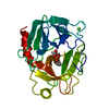
| ||||||||
|---|---|---|---|---|---|---|---|---|---|
| 1 |
| ||||||||
| Unit cell |
|
-Components
| #1: Protein | Mass: 23446.346 Da / Num. of mol.: 1 / Source method: isolated from a natural source / Source: (natural) |
|---|---|
| #2: Chemical | ChemComp-CA /   Mass: 40.078 Da / Num. of mol.: 1 / Source method: obtained synthetically / Formula: Ca Mass: 40.078 Da / Num. of mol.: 1 / Source method: obtained synthetically / Formula: Ca |
| #3: Water | ChemComp-HOH /  Mass: 18.015 Da / Num. of mol.: 211 / Source method: isolated from a natural source / Formula: H2O Mass: 18.015 Da / Num. of mol.: 211 / Source method: isolated from a natural source / Formula: H2O |
| Has protein modification | Y |
-Experimental details
-Experiment
| Experiment | Method: X-RAY DIFFRACTION |
|---|
- Sample preparation
| Crystal | Density Matthews: 2.31 Å3/Da / Density % sol: 46.79 % | ||||||||||||||||||||
|---|---|---|---|---|---|---|---|---|---|---|---|---|---|---|---|---|---|---|---|---|---|
| Crystal grow | *PLUS pH: 7 / Method: unknown | ||||||||||||||||||||
| Components of the solutions | *PLUS
|
-Data collection
| Radiation | Scattering type: x-ray |
|---|---|
| Radiation wavelength | Relative weight: 1 |
- Processing
| Software | Name: PROLSQ / Classification: refinement | |||||||||||||||||||||||||||||||||||||||||||||||||||||||||||||||
|---|---|---|---|---|---|---|---|---|---|---|---|---|---|---|---|---|---|---|---|---|---|---|---|---|---|---|---|---|---|---|---|---|---|---|---|---|---|---|---|---|---|---|---|---|---|---|---|---|---|---|---|---|---|---|---|---|---|---|---|---|---|---|---|---|
| Refinement | Resolution: 1.34→7 Å Details: THE TRYPSIN IN THE CRYSTAL CONTAINS ABOUT 50 PER-CENT ALPHA-TRYPSIN WHICH IS AUTOLYTICALLY CLEAVED BETWEEN LYS 145 AND SER 146. THE DENSITY IN THE MAP AT THIS POINT IS WEAK BUT APPEARS TO ...Details: THE TRYPSIN IN THE CRYSTAL CONTAINS ABOUT 50 PER-CENT ALPHA-TRYPSIN WHICH IS AUTOLYTICALLY CLEAVED BETWEEN LYS 145 AND SER 146. THE DENSITY IN THE MAP AT THIS POINT IS WEAK BUT APPEARS TO ARISE FROM THE UNCLEAVED COMPONENT.
| |||||||||||||||||||||||||||||||||||||||||||||||||||||||||||||||
| Displacement parameters | Biso mean: 16 Å2 | |||||||||||||||||||||||||||||||||||||||||||||||||||||||||||||||
| Refinement step | Cycle: LAST / Resolution: 1.34→7 Å
| |||||||||||||||||||||||||||||||||||||||||||||||||||||||||||||||
| Refine LS restraints |
| |||||||||||||||||||||||||||||||||||||||||||||||||||||||||||||||
| Refinement | *PLUS Rfactor obs: 0.152 | |||||||||||||||||||||||||||||||||||||||||||||||||||||||||||||||
| Solvent computation | *PLUS | |||||||||||||||||||||||||||||||||||||||||||||||||||||||||||||||
| Displacement parameters | *PLUS |
