 Movie
Movie Controller

[English] 日本語
 Yorodumi
- PDB-1ntp: USE OF THE NEUTRON DIFFRACTION H/D EXCHANGE TECHNIQUE TO DETERMIN... -
+ Open data

- Basic information
| Entry | Database: PDB / ID: 1ntp | ||||||
|---|---|---|---|---|---|---|---|
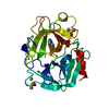
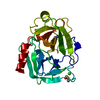
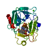
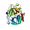
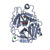
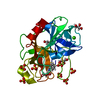
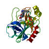
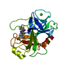
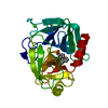
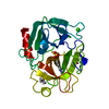
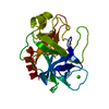
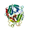
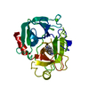
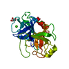
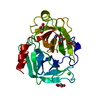
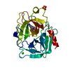
| Title | USE OF THE NEUTRON DIFFRACTION H/D EXCHANGE TECHNIQUE TO DETERMINE THE CONFORMATIONAL DYNAMICS OF TRYPSIN | ||||||
 Components Components | BETA-TRYPSIN | ||||||
 Keywords Keywords | HYDROLASE (SERINE PROTEINASE) | ||||||
| Function / homology |  Function and homology information Function and homology informationtrypsin / serpin family protein binding / serine protease inhibitor complex / digestion / endopeptidase activity / serine-type endopeptidase activity / proteolysis / : / metal ion binding Similarity search - Function | ||||||
| Biological species |  | ||||||
| Method | NEUTRON DIFFRACTION / Resolution: 1.8 Å | ||||||
 Authors Authors | Kossiakoff, A.A. | ||||||
 Citation Citation | Journal: Basic Life Sci. / Year: 1984 Title: Use of the neutron diffraction--H/D exchange technique to determine the conformational dynamics of trypsin Authors: Kossiakoff, A.A. #1: Journal: Nature / Year: 1982Title: Protein Dynamics Investigated by the Neutron Diffraction-Hydrogen Exchange Technique Authors: Kossiakoff, A.A. #2: Journal: Biochemistry / Year: 1981Title: Direct Determination of the Protonation States of Aspartic Acid-102 and Histidine-57 in the Tetrahedral Intermediate of the Serine Proteases. Neutron Structure of Trypsin Authors: Kossiakoff, A.A. / Spencer, S.A. #3: Journal: Nature / Year: 1980Title: Neutron Diffraction Identifies His 57 as the Catalytic Base in Trypsin Authors: Kossiakoff, A.A. / Spencer, S.A. #4: Journal: Acta Crystallogr.,Sect.B / Year: 1979Title: The Accuracy of Refined Protein Structures, Comparison of Two Independently Refined Models of Bovine Trypsin Authors: Chambers, J.L. / Stroud, R.M. #5: Journal: Acta Crystallogr.,Sect.B / Year: 1977Title: Difference-Fourier Refinement of the Structure of Dip-Trypsin at 1.5 Angstroms Using a Minicomputer Technique Authors: Chambers, J.L. / Stroud, R.M. #6: Journal: Proteases and Biological Control / Year: 1975Title: Structure-Function Relationships in the Serine Proteases Authors: Stroud, R.M. / Krieger, M. / Koeppeii, R.E. / Kossiakoff, A.A. / Chambers, J.L. #7: Journal: Biochem.Biophys.Res.Commun. / Year: 1974Title: Silver Ion Inhibition of Serine Proteases, Crystallographic Study of Silver-Trypsin Authors: Chambers, J.L. / Christoph, G.G. / Krieger, M. / Kay, L. / Stroud, R.M. #8: Journal: J.Mol.Biol. / Year: 1974Title: The Structure of Bovine Trypsin,Electron Density Maps of the Inhibited Enzyme at 5 Angstroms and at 2.7 Angstroms Resolution Authors: Stroud, R.M. / Kay, L.M. / Dickerson, R.E. #9: Journal: J.Mol.Biol. / Year: 1974Title: Structure and Specific Binding of Trypsin, Comparison of Inhibited Derivatives and a Model for Substrate Binding Authors: Krieger, M. / Kay, L.M. / Stroud, R.M. #10: Journal: Cold Spring Harbor Symp.Quant.Biol. / Year: 1972Title: The Crystal and Molecular Structure of Dip-Inhibited Bovine Trypsin at 2.7 Angstroms Resolution Authors: Stroud, R.M. / Kay, L.M. / Dickerson, R.E. | ||||||
| History |
|
- Structure visualization
| Structure viewer | Molecule: MolmilJmol/JSmol |
|---|
- Downloads & links
-Download
| PDBx/mmCIF format | 1ntp.cif.gz | 83.2 KB | Display | PDBx/mmCIF format |
|---|---|---|---|---|
| PDB format | pdb1ntp.ent.gz | 63.6 KB | Display | PDB format |
| PDBx/mmJSON format | 1ntp.json.gz | Tree view | PDBx/mmJSON format | |
| Others |  Other downloads Other downloads |
-Validation report
| Arichive directory | https://data.pdbj.org/pub/pdb/validation_reports/nt/1ntpftp://data.pdbj.org/pub/pdb/validation_reports/nt/1ntp | HTTPS FTP |
|---|
-Related structure data
| Similar structure data |
|---|


-Links
 PDBj
PDBj

- Assembly
| Deposited unit | 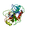
| ||||||||
|---|---|---|---|---|---|---|---|---|---|
| 1 |
| ||||||||
| Unit cell |
| ||||||||
| Atom site foot note | 1: SEE REMARK 4. 2: AN OCCUPANCY OF 0.0 INDICATES THAT NO SIGNIFICANT ELECTRON DENSITY WAS FOUND IN THE FINAL FOURIER MAP. |
-Components
| #1: Protein | Mass: 23327.242 Da / Num. of mol.: 1 Source method: isolated from a genetically manipulated source Source: (gene. exp.) |
|---|---|
| #2: Chemical | ChemComp-ISP /   Mass: 140.075 Da / Num. of mol.: 1 / Source method: obtained synthetically / Formula: C3H9O4P Mass: 140.075 Da / Num. of mol.: 1 / Source method: obtained synthetically / Formula: C3H9O4P |
| Has protein modification | Y |
| Nonpolymer details | THE ENZYME IS INHIBITED BY A MONOISOPROPYLPHOSPHORYL DERIVATIVE. THE REFINED STRUCTURE IN THIS ...THE ENZYME IS INHIBITED BY A MONOISOPRO |
-Experimental details
-Experiment
| Experiment | Method: NEUTRON DIFFRACTION |
|---|
-Data collection
| Radiation | Scattering type: neutron |
|---|---|
| Radiation wavelength | Relative weight: 1 |
- Processing
| Refinement | Rfactor Rwork: 0.187 / Highest resolution: 1.8 Å | ||||||||||||||||||||||||||||||||||||||||||||||||||||||||||||
|---|---|---|---|---|---|---|---|---|---|---|---|---|---|---|---|---|---|---|---|---|---|---|---|---|---|---|---|---|---|---|---|---|---|---|---|---|---|---|---|---|---|---|---|---|---|---|---|---|---|---|---|---|---|---|---|---|---|---|---|---|---|
| Refinement step | Cycle: LAST / Highest resolution: 1.8 Å
| ||||||||||||||||||||||||||||||||||||||||||||||||||||||||||||
| Refine LS restraints |
|
