 Movie
Movie Controller

[English] 日本語
 Yorodumi
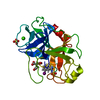
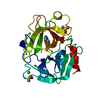
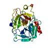
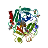
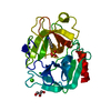
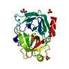
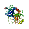
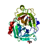
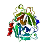
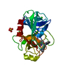
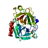
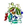
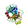
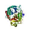
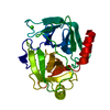
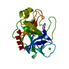
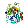
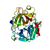
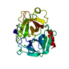
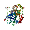
- PDB-1gi2: A NOVEL SERINE PROTEASE INHIBITION MOTIF INVOLVING A MULTI-CENTER... -
+ Open data

- Basic information
| Entry | Database: PDB / ID: 1gi2 | ||||||
|---|---|---|---|---|---|---|---|
| Title | A NOVEL SERINE PROTEASE INHIBITION MOTIF INVOLVING A MULTI-CENTERED SHORT HYDROGEN BONDING NETWORK AT THE ACTIVE SITE | ||||||
 Components Components | BETA-TRYPSIN | ||||||
 Keywords Keywords | HYDROLASE / three-centered / very short hydrogen bond / oxyanion hole water / shift of pKa of His57 / structure-based drug design / specificity / urokinase / trypsin / thrombin / Zn+2-mediated inhibition | ||||||
| Function / homology |  Function and homology information Function and homology informationtrypsin / serpin family protein binding / serine protease inhibitor complex / digestion / endopeptidase activity / serine-type endopeptidase activity / proteolysis / : / metal ion binding Similarity search - Function | ||||||
| Biological species |  | ||||||
| Method |  X-RAY DIFFRACTION / FOURIER SYNTHESIS / Resolution: 1.38 Å X-RAY DIFFRACTION / FOURIER SYNTHESIS / Resolution: 1.38 Å | ||||||
 Authors Authors | Katz, B.A. / Elrod, K. / Luong, C. / Rice, M. / Mackman, R.L. / Sprengeler, P.A. / Spencer, J. / Hatayte, J. / Janc, J. / Link, J. ...Katz, B.A. / Elrod, K. / Luong, C. / Rice, M. / Mackman, R.L. / Sprengeler, P.A. / Spencer, J. / Hatayte, J. / Janc, J. / Link, J. / Litvak, J. / Rai, R. / Rice, K. / Sideris, S. / Verner, E. / Young, W. | ||||||
 Citation Citation | Journal: J.Mol.Biol. / Year: 2001 Title: A novel serine protease inhibition motif involving a multi-centered short hydrogen bonding network at the active site. Authors: Katz, B.A. / Elrod, K. / Luong, C. / Rice, M.J. / Mackman, R.L. / Sprengeler, P.A. / Spencer, J. / Hataye, J. / Janc, J. / Link, J. / Litvak, J. / Rai, R. / Rice, K. / Sideris, S. / Verner, E. / Young, W. | ||||||
| History |
|
- Structure visualization
| Structure viewer | Molecule: MolmilJmol/JSmol |
|---|
- Downloads & links
-Download
| PDBx/mmCIF format | 1gi2.cif.gz | 110.7 KB | Display | PDBx/mmCIF format |
|---|---|---|---|---|
| PDB format | pdb1gi2.ent.gz | 87.4 KB | Display | PDB format |
| PDBx/mmJSON format | 1gi2.json.gz | Tree view | PDBx/mmJSON format | |
| Others |  Other downloads Other downloads |
-Validation report
| Arichive directory | https://data.pdbj.org/pub/pdb/validation_reports/gi/1gi2ftp://data.pdbj.org/pub/pdb/validation_reports/gi/1gi2 | HTTPS FTP |
|---|
-Related structure data
| Related structure data |  1ghvC  1ghwC  1ghxC  1ghyC 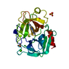 1ghzC 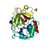 1gi0C 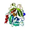 1gi1C 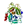 1gi3C 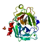 1gi4C  1gi5C 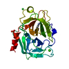 1gi6C  1gi7C  1gi8C  1gi9C C: citing same article ( |
|---|---|
| Similar structure data |

-Links
 PDBj
PDBj


- Assembly
| Deposited unit | 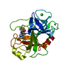
| ||||||||
|---|---|---|---|---|---|---|---|---|---|
| 1 |
| ||||||||
| Unit cell |
|
-Components
| #1: Protein | Mass: 23324.287 Da / Num. of mol.: 1 / Source method: isolated from a natural source / Source: (natural) |
|---|---|
| #2: Chemical | ChemComp-CA /   Mass: 40.078 Da / Num. of mol.: 1 / Source method: obtained synthetically / Formula: Ca Mass: 40.078 Da / Num. of mol.: 1 / Source method: obtained synthetically / Formula: Ca |
| #3: Chemical | ChemComp-SO4 /   Mass: 96.063 Da / Num. of mol.: 1 / Source method: obtained synthetically / Formula: SO4 Mass: 96.063 Da / Num. of mol.: 1 / Source method: obtained synthetically / Formula: SO4 |
| #4: Chemical | ChemComp-122 / 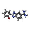  Mass: 253.279 Da / Num. of mol.: 1 / Source method: obtained synthetically / Formula: C14H13N4O Mass: 253.279 Da / Num. of mol.: 1 / Source method: obtained synthetically / Formula: C14H13N4O |
| #5: Water | ChemComp-HOH /  Mass: 18.015 Da / Num. of mol.: 269 / Source method: isolated from a natural source / Formula: H2O Mass: 18.015 Da / Num. of mol.: 269 / Source method: isolated from a natural source / Formula: H2O |
| Has protein modification | Y |
-Experimental details
-Experiment
| Experiment | Method: X-RAY DIFFRACTION / Number of used crystals: 1 |
|---|
- Sample preparation
| Crystal | Density Matthews: 2.32 Å3/Da / Density % sol: 47.06 % |
|---|---|
| Crystal grow | Temperature: 298 K / Method: vapor diffusion / pH: 8.1 Details: magnesium sulfate soak at target pH (8.1). vapor diffusion at 298 K, pH 8.10 |
-Data collection
| Diffraction | Mean temperature: 298 K |
|---|---|
| Diffraction source | Source: ROTATING ANODE / Type: RIGAKU RU200 / Wavelength: 1.5418 |
| Detector | Type: RIGAKU RAXIS IV / Detector: IMAGE PLATE / Date: Jul 25, 1999 |
| Radiation | Protocol: SINGLE WAVELENGTH / Monochromatic (M) / Laue (L): M / Scattering type: x-ray |
| Radiation wavelength | Wavelength: 1.5418 Å / Relative weight: 1 |
| Reflection | Resolution: 1.32→67.43 Å / Num. all: 51781 / Num. obs: 36102 / % possible obs: 69.7 % / Observed criterion σ(I): 0.8 / Redundancy: 2.8 % / Rmerge(I) obs: 0.088 / Net I/σ(I): 7.9 |
| Reflection shell | Resolution: 1.38→1.44 Å / Rmerge(I) obs: 0.275 / Num. unique all: 1889 / % possible all: 34.1 |
- Processing
| Software |
| ||||||||||||||||||||
|---|---|---|---|---|---|---|---|---|---|---|---|---|---|---|---|---|---|---|---|---|---|
| Refinement | Method to determine structure: FOURIER SYNTHESIS / Resolution: 1.38→7 Å / σ(F): 1.7 / Stereochemistry target values: X-PLOR force field Details: Residues simultaneously refined in two or more conformations are: Val53, Ser61, Ser84, Ser86, Leu99, Met104, Ser113, Ser116, Arg117, Ser127 Gln135, Ser146, Asp165, Ser166, Ser167, Gln175, ...Details: Residues simultaneously refined in two or more conformations are: Val53, Ser61, Ser84, Ser86, Leu99, Met104, Ser113, Ser116, Arg117, Ser127 Gln135, Ser146, Asp165, Ser166, Ser167, Gln175, Ser217, Ser236, Ser244. Note that HOH383 makes short H-bonds to OgSer195 and O6' of the inhibitor Disordered waters are: HOH379 which is close to sulfate_380; HOH425 which is close to HOH426; HOH461 which is close to a symmetry-related equivalent of HOH462; HOH604 which is close to HOH605; HIS91 is MONOPROTONATED ON THE EPSILON NITROGEN. His57 is doubly protonatd.
| ||||||||||||||||||||
| Refinement step | Cycle: LAST / Resolution: 1.38→7 Å
| ||||||||||||||||||||
| Refine LS restraints |
|
