 Movie
Movie Controller

[English] 日本語
 Yorodumi
- PDB-2tga: ON THE DISORDERED ACTIVATION DOMAIN IN TRYPSINOGEN. CHEMICAL LABE... -
+ Open data

- Basic information
| Entry | Database: PDB / ID: 2tga | |||||||||
|---|---|---|---|---|---|---|---|---|---|---|
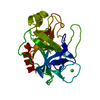
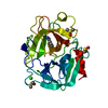
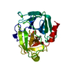
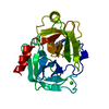
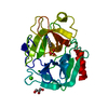
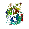
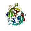
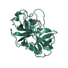
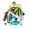
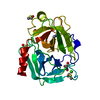
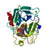
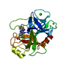
| Title | ON THE DISORDERED ACTIVATION DOMAIN IN TRYPSINOGEN. CHEMICAL LABELLING AND LOW-TEMPERATURE CRYSTALLOGRAPHY | |||||||||
 Components Components | TRYPSINOGEN | |||||||||
 Keywords Keywords | HYDROLASE ZYMOGEN (SERINE PROTEINASE) | |||||||||
| Function / homology |  Function and homology information Function and homology informationtrypsin / serpin family protein binding / serine protease inhibitor complex / digestion / endopeptidase activity / serine-type endopeptidase activity / proteolysis / : / metal ion binding Similarity search - Function | |||||||||
| Biological species |  | |||||||||
| Method |  X-RAY DIFFRACTION / Resolution: 1.8 Å X-RAY DIFFRACTION / Resolution: 1.8 Å | |||||||||
 Authors Authors | Walter, J. / Steigemann, W. / Singh, T.P. / Bartunik, H. / Bode, W. / Huber, R. | |||||||||
 Citation Citation | Journal: Acta Crystallogr.,Sect.B / Year: 1982 Title: On the Disordered Activation Domain in Trypsinogen. Chemical Labelling and Low-Temperature Crystallography Authors: Walter, J. / Steigemann, W. / Singh, T.P. / Bartunik, H. / Bode, W. / Huber, R. #1: Journal: Acta Crystallogr.,Sect.B / Year: 1983Title: The Geometry of the Reactive Site and of the Peptide Groups in Trypsin, Trypsinogen and its Complexes with Inhibitors Authors: Marquart, M. / Walter, J. / Deisenhofer, J. / Bode, W. / Huber, R. #2: Journal: Acta Crystallogr.,Sect.B / Year: 1980Title: Low-Temperature Protein Crystallography. Effect on Flexibility, Temperature Factor, Mosaic Spread, Extinction and Diffuse Scattering in Two Examples. Bovine Trypsinogen and Fc Fragment Authors: Singh, T.P. / Bode, W. / Huber, R. #3: Journal: Acc.Chem.Res. / Year: 1978Title: Structural Basis of the Activation and Action of Trypsin Authors: Huber, R. / Bode, W. #4: Journal: FEBS Lett. / Year: 1978Title: Crystal Structure Analysis and Refinement of Two Variants of Trigonal Trypsinogen Authors: Bode, W. / Huber, R. #5: Journal: J.Mol.Biol. / Year: 1977Title: Crystal Structure of Bovine Trypsinogen at 1.8 Angstroms Resolution. II. Crystallographic Refinement, Refined Crystal Structure and Comparison with Bovine Trypsin Authors: Fehlhammer, H. / Bode, W. / Huber, R. #6: Journal: J.Mol.Biol. / Year: 1976Title: Crystal Structure of Bovine Trypsinogen at 1.8 Angstroms Resolution. I. Data Collection, Application of Patterson Search Techniques and Preliminary Structural Interpretation Authors: Bode, W. / Fehlhammer, H. / Huber, R. | |||||||||
| History |
|
- Structure visualization
| Structure viewer | Molecule: MolmilJmol/JSmol |
|---|
- Downloads & links
-Download
| PDBx/mmCIF format | 2tga.cif.gz | 53.4 KB | Display | PDBx/mmCIF format |
|---|---|---|---|---|
| PDB format | pdb2tga.ent.gz | 38.8 KB | Display | PDB format |
| PDBx/mmJSON format | 2tga.json.gz | Tree view | PDBx/mmJSON format | |
| Others |  Other downloads Other downloads |
-Validation report
| Arichive directory | https://data.pdbj.org/pub/pdb/validation_reports/tg/2tgaftp://data.pdbj.org/pub/pdb/validation_reports/tg/2tga | HTTPS FTP |
|---|
-Related structure data
| Similar structure data |
|---|


-Links
 PDBj
PDBj

- Assembly
| Deposited unit | 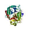
| ||||||||
|---|---|---|---|---|---|---|---|---|---|
| 1 |
| ||||||||
| Unit cell |
| ||||||||
| Atom site foot note | 1: SEE REMARKS 4 AND 6. |
-Components
| #1: Protein | Mass: 24012.953 Da / Num. of mol.: 1 Source method: isolated from a genetically manipulated source Source: (gene. exp.) |
|---|---|
| #2: Chemical | ChemComp-CA /   Mass: 40.078 Da / Num. of mol.: 1 / Source method: obtained synthetically / Formula: Ca Mass: 40.078 Da / Num. of mol.: 1 / Source method: obtained synthetically / Formula: Ca |
| #3: Water | ChemComp-HOH /  Mass: 18.015 Da / Num. of mol.: 93 / Source method: isolated from a natural source / Formula: H2O Mass: 18.015 Da / Num. of mol.: 93 / Source method: isolated from a natural source / Formula: H2O |
| Has protein modification | Y |
| Sequence details | THE 229 AMINO ACIDS OF TRYPSINOGE |
-Experimental details
-Experiment
| Experiment | Method: X-RAY DIFFRACTION |
|---|
- Sample preparation
| Crystal | Density Matthews: 2 Å3/Da / Density % sol: 38.38 % | |||||||||||||||||||||||||||||||||||
|---|---|---|---|---|---|---|---|---|---|---|---|---|---|---|---|---|---|---|---|---|---|---|---|---|---|---|---|---|---|---|---|---|---|---|---|---|
| Crystal grow | *PLUS pH: 7 / Method: vapor diffusion | |||||||||||||||||||||||||||||||||||
| Components of the solutions | *PLUS
|
-Data collection
| Reflection | *PLUS Highest resolution: 1.8 Å / Num. obs: 13952 / % possible obs: 75.4 % / Biso Wilson estimate: 12.4 Å2 |
|---|
- Processing
| Refinement | Resolution: 1.8→6.5 Å / Rfactor Rwork: 0.197 Details: THERE ARE FOUR *FLEXIBLE* SEGMENTS FOR WHICH THERE IS NO SIGNIFICANT ELECTRON DENSITY IN THE MAP. THESE ARE 1. THREE CLOSELY INTERDIGITATING CHAIN SEGMENTS FORMING THE ACTIVATION DOMAIN GLY ...Details: THERE ARE FOUR *FLEXIBLE* SEGMENTS FOR WHICH THERE IS NO SIGNIFICANT ELECTRON DENSITY IN THE MAP. THESE ARE 1. THREE CLOSELY INTERDIGITATING CHAIN SEGMENTS FORMING THE ACTIVATION DOMAIN GLY 142 - PRO 152 GLY 184A - GLY 193 GLY 216 - ASN 223 2. THE N-TERMINUS FROM VAL 10 THROUGH GLY 18 (THIS DATA ENTRY CONTAINS NO COORDINATES FOR VAL 10 THROUGH LYS 15) | ||||||||||||
|---|---|---|---|---|---|---|---|---|---|---|---|---|---|
| Refinement step | Cycle: LAST / Resolution: 1.8→6.5 Å
| ||||||||||||
| Refinement | *PLUS Highest resolution: 1.8 Å / Lowest resolution: 6.5 Å / Rfactor obs: 0.197 | ||||||||||||
| Solvent computation | *PLUS | ||||||||||||
| Displacement parameters | *PLUS |
