 Movie
Movie Controller

[English] 日本語
 Yorodumi
- PDB-2tld: CRYSTAL STRUCTURE OF AN ENGINEERED SUBTILISIN INHIBITOR COMPLEXED... -
+ Open data

- Basic information
| Entry | Database: PDB / ID: 2tld | ||||||
|---|---|---|---|---|---|---|---|
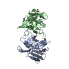
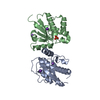
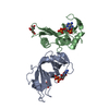
| Title | CRYSTAL STRUCTURE OF AN ENGINEERED SUBTILISIN INHIBITOR COMPLEXED WITH BOVINE TRYPSIN | ||||||
 Components Components |
| ||||||
 Keywords Keywords | HYDROLASE/HYDROLASE INHIBITOR / PROTEINASE / HYDROLASE-HYDROLASE INHIBITOR COMPLEX / TRYPSIN) | ||||||
| Function / homology |  Function and homology information Function and homology informationtrypsin / serpin family protein binding / serine protease inhibitor complex / digestion / serine-type endopeptidase inhibitor activity / endopeptidase activity / serine-type endopeptidase activity / proteolysis / : / extracellular region / metal ion binding Similarity search - Function | ||||||
| Biological species |  | ||||||
| Method |  X-RAY DIFFRACTION / Resolution: 2.6 Å X-RAY DIFFRACTION / Resolution: 2.6 Å | ||||||
 Authors Authors | Mitsui, Y. / Takeuchi, Y. / Nonaka, T. / Nakamura, K.T. | ||||||
 Citation Citation | Journal: Proc.Natl.Acad.Sci.USA / Year: 1992 Title: Crystal structure of an engineered subtilisin inhibitor complexed with bovine trypsin. Authors: Takeuchi, Y. / Nonaka, T. / Nakamura, K.T. / Kojima, S. / Miura, K. / Mitsui, Y. #1: Journal: J.Mol.Biol. / Year: 1991Title: Refined Crystal Structure of the Complex of Subtilisin Bpn' and Streptomyces Subtilisin Inhibitor at 1.8 Angstroms Resolution Authors: Takeuchi, Y. / Satow, Y. / Nakamura, K.T. / Mitsui, Y. | ||||||
| History |
|
- Structure visualization
| Structure viewer | Molecule: MolmilJmol/JSmol |
|---|
- Downloads & links
-Download
| PDBx/mmCIF format | 2tld.cif.gz | 24.5 KB | Display | PDBx/mmCIF format |
|---|---|---|---|---|
| PDB format | pdb2tld.ent.gz | 11.9 KB | Display | PDB format |
| PDBx/mmJSON format | 2tld.json.gz | Tree view | PDBx/mmJSON format | |
| Others |  Other downloads Other downloads |
-Validation report
| Arichive directory | https://data.pdbj.org/pub/pdb/validation_reports/tl/2tldftp://data.pdbj.org/pub/pdb/validation_reports/tl/2tld | HTTPS FTP |
|---|
-Related structure data
| Related structure data | |
|---|---|
| Similar structure data |

-Links
 PDBj
PDBj

- Assembly
| Deposited unit | 
| ||||||||
|---|---|---|---|---|---|---|---|---|---|
| 1 |
| ||||||||
| Unit cell |
| ||||||||
| Atom site foot note | 1: PRO I 37 IS A CIS PROLINE. | ||||||||
| Details | SSI IS A DIMERIC MOLECULE (I2) CONSISTING OF TWO IDENTICAL SUBUNITS. IT BINDS TWO TRYPSIN MOLECULES (2E) TO FORM A DIMERIC COMPLEX E2I2. THE CRYSTALLOGRAPHIC ASYMMETRIC UNIT CORRESPONDS TO HALF THE COMPLEX MOLECULE (EI). IN THIS ENTRY COORDINATES FOR ALPHA-CARBON ATOMS ARE PROVIDED FOR ONE CHAIN OF TRYPSIN USING THE CHAIN IDENTIFIER *E* AND FOR ONE CHAIN OF MODIFIED SSI USING THE CHAIN IDENTIFIER *I*. COORDINATES FOR THE OTHER EI COMPLEX CAN BE GENERATED FROM THE COORDINATES IN THIS ENTRY USING THE FOLLOWING TRANSFORMATION: -1.0 0.0 0.0 0.0 -1.0 0.0 0.0 0.0 1.0 |
-Components
| #1: Protein | Mass: 22903.805 Da / Num. of mol.: 1 Source method: isolated from a genetically manipulated source Source: (gene. exp.) |
|---|---|
| #2: Protein | Mass: 11135.486 Da / Num. of mol.: 1 Source method: isolated from a genetically manipulated source References: UniProt: P01006 |
-Experimental details
-Experiment
| Experiment | Method: X-RAY DIFFRACTION |
|---|
- Sample preparation
| Crystal | Density Matthews: 3.06 Å3/Da / Density % sol: 59.77 % | ||||||||||||||||||||||||||||||
|---|---|---|---|---|---|---|---|---|---|---|---|---|---|---|---|---|---|---|---|---|---|---|---|---|---|---|---|---|---|---|---|
| Crystal grow | *PLUS pH: 7 / Method: vapor diffusion, hanging drop | ||||||||||||||||||||||||||||||
| Components of the solutions | *PLUS
|
-Data collection
| Radiation | Scattering type: x-ray |
|---|---|
| Radiation wavelength | Relative weight: 1 |
| Reflection | *PLUS Highest resolution: 2.6 Å / Num. obs: 7579 / Observed criterion σ(I): 2 / Num. measured all: 23106 / Rmerge(I) obs: 0.063 |
- Processing
| Software | Name: X-PLOR / Classification: refinement | ||||||||||||||||||||||||||||||||||||||||||||||||||||||||||||
|---|---|---|---|---|---|---|---|---|---|---|---|---|---|---|---|---|---|---|---|---|---|---|---|---|---|---|---|---|---|---|---|---|---|---|---|---|---|---|---|---|---|---|---|---|---|---|---|---|---|---|---|---|---|---|---|---|---|---|---|---|---|
| Refinement | Resolution: 2.6→8 Å /
| ||||||||||||||||||||||||||||||||||||||||||||||||||||||||||||
| Refinement step | Cycle: LAST / Resolution: 2.6→8 Å
| ||||||||||||||||||||||||||||||||||||||||||||||||||||||||||||
| Refine LS restraints |
| ||||||||||||||||||||||||||||||||||||||||||||||||||||||||||||
| Software | *PLUS Name: X-PLOR / Classification: refinement | ||||||||||||||||||||||||||||||||||||||||||||||||||||||||||||
| Refinement | *PLUS Highest resolution: 2.6 Å / Lowest resolution: 8 Å / Num. reflection all: 6166 / Rfactor obs: 0.173 | ||||||||||||||||||||||||||||||||||||||||||||||||||||||||||||
| Solvent computation | *PLUS | ||||||||||||||||||||||||||||||||||||||||||||||||||||||||||||
| Displacement parameters | *PLUS | ||||||||||||||||||||||||||||||||||||||||||||||||||||||||||||
| Refine LS restraints | *PLUS
|
