 Movie
Movie Controller

[English] 日本語
 Yorodumi
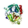
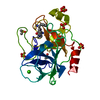
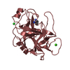
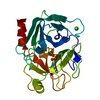
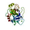
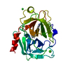
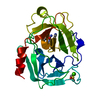
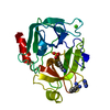
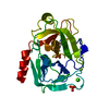
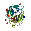
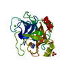
- PDB-2tgd: LACK OF THE TRANSITION STATE STABILIZATION SITE IS A FACTOR IN TH... -
+ Open data

- Basic information
| Entry | Database: PDB / ID: 2tgd | ||||||
|---|---|---|---|---|---|---|---|
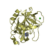
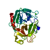
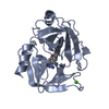
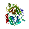
| Title | LACK OF THE TRANSITION STATE STABILIZATION SITE IS A FACTOR IN THE INACTIVITY OF TRYPSINOGEN, A SERINE PROTEASE ZYMOGEN. STRUCTURE OF DFP INHIBITED BOVINE TRYPSINOGEN AT 2.1 ANGSTROMS RESOLUTION | ||||||
 Components Components | TRYPSINOGEN | ||||||
 Keywords Keywords | HYDROLASE ZYMOGEN (SERINE PROTEINASE) | ||||||
| Function / homology |  Function and homology information Function and homology informationtrypsin / serpin family protein binding / serine protease inhibitor complex / digestion / endopeptidase activity / serine-type endopeptidase activity / proteolysis / : / metal ion binding Similarity search - Function | ||||||
| Biological species |  | ||||||
| Method |  X-RAY DIFFRACTION / Resolution: 2.1 Å X-RAY DIFFRACTION / Resolution: 2.1 Å | ||||||
 Authors Authors | Jones, M.O. / Stroud, R.M. | ||||||
 Citation Citation | Journal: To be Published Title: Lack of the Transition State Stabilization Site is a Factor in the Inactivity of Trypsinogen, a Serine Protease Zymogen. Structure of Dfp Inhibited Bovine Trypsinogen at 2.1 Angstroms Resolution Authors: Jones, M.O. / Stroud, R.M. #1: Journal: Biochemistry / Year: 1977Title: Structure of Bovine Trypsinogen at 1.9 Angstroms Resolution Authors: Kossiakoff, A.A. / Chambers, J.L. / Kay, L.M. / Stroud, R.M. #2: Journal: Annu.Rev.Biophys.Bioeng. / Year: 1977Title: Mechanisms of Zymogen Activation Authors: Stroud, R.M. / Kossiakoff, A.A. / Chambers, J.L. | ||||||
| History |
|
- Structure visualization
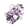
| Structure viewer | Molecule: MolmilJmol/JSmol |
|---|
- Downloads & links
-Download
| PDBx/mmCIF format | 2tgd.cif.gz | 54.3 KB | Display | PDBx/mmCIF format |
|---|---|---|---|---|
| PDB format | pdb2tgd.ent.gz | 35.1 KB | Display | PDB format |
| PDBx/mmJSON format | 2tgd.json.gz | Tree view | PDBx/mmJSON format | |
| Others |  Other downloads Other downloads |
-Validation report
| Arichive directory | https://data.pdbj.org/pub/pdb/validation_reports/tg/2tgdftp://data.pdbj.org/pub/pdb/validation_reports/tg/2tgd | HTTPS FTP |
|---|
-Related structure data
| Similar structure data |
|---|


-Links
 PDBj
PDBj

- Assembly
| Deposited unit | 
| ||||||||
|---|---|---|---|---|---|---|---|---|---|
| 1 |
| ||||||||
| Unit cell |
| ||||||||
| Atom site foot note | 1: SEE REMARK 6. |
-Components
| #1: Protein | Mass: 24012.953 Da / Num. of mol.: 1 Source method: isolated from a genetically manipulated source Source: (gene. exp.) |
|---|---|
| #2: Chemical | ChemComp-CA /   Mass: 40.078 Da / Num. of mol.: 1 / Source method: obtained synthetically / Formula: Ca Mass: 40.078 Da / Num. of mol.: 1 / Source method: obtained synthetically / Formula: Ca |
| #3: Chemical | ChemComp-DFP /   Mass: 166.155 Da / Num. of mol.: 1 / Source method: obtained synthetically / Formula: C6H15O3P Mass: 166.155 Da / Num. of mol.: 1 / Source method: obtained synthetically / Formula: C6H15O3P |
| #4: Water | ChemComp-HOH /  Mass: 18.015 Da / Num. of mol.: 73 / Source method: isolated from a natural source / Formula: H2O Mass: 18.015 Da / Num. of mol.: 73 / Source method: isolated from a natural source / Formula: H2O |
| Has protein modification | Y |
-Experimental details
-Experiment
| Experiment | Method: X-RAY DIFFRACTION |
|---|
- Sample preparation
| Crystal | Density Matthews: 2 Å3/Da / Density % sol: 38.42 % |
|---|
- Processing
| Refinement | Rfactor Rwork: 0.182 / Highest resolution: 2.1 Å | ||||||||||||||||||||||||||||||||||||||||||||||||||||||||||||
|---|---|---|---|---|---|---|---|---|---|---|---|---|---|---|---|---|---|---|---|---|---|---|---|---|---|---|---|---|---|---|---|---|---|---|---|---|---|---|---|---|---|---|---|---|---|---|---|---|---|---|---|---|---|---|---|---|---|---|---|---|---|
| Refinement step | Cycle: LAST / Highest resolution: 2.1 Å
| ||||||||||||||||||||||||||||||||||||||||||||||||||||||||||||
| Refine LS restraints |
|
