 Movie
Movie Controller

[English] 日本語
 Yorodumi
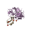
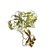
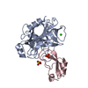
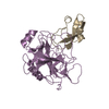
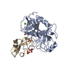
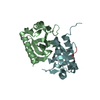
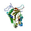
- PDB-3btf: THE CRYSTAL STRUCTURES OF THE COMPLEXES BETWEEN BOVINE BETA-TRYPS... -
+ Open data

- Basic information
| Entry | Database: PDB / ID: 3btf | ||||||
|---|---|---|---|---|---|---|---|
| Title | THE CRYSTAL STRUCTURES OF THE COMPLEXES BETWEEN BOVINE BETA-TRYPSIN AND TEN P1 VARIANTS OF BPTI. | ||||||
 Components Components |
| ||||||
 Keywords Keywords | HYDROLASE/HYDROLASE INHIBITOR / TRYPSIN / BPTI / SERINE PROTEINASE / INHIBITOR / HYDROLASE-HYDROLASE INHIBITOR COMPLEX | ||||||
| Function / homology |  Function and homology information Function and homology informationsulfate binding / negative regulation of platelet aggregation / zymogen binding / potassium channel inhibitor activity / molecular function inhibitor activity / negative regulation of thrombin-activated receptor signaling pathway / trypsin / serpin family protein binding / serine protease inhibitor complex / digestion ...sulfate binding / negative regulation of platelet aggregation / zymogen binding / potassium channel inhibitor activity / molecular function inhibitor activity / negative regulation of thrombin-activated receptor signaling pathway / trypsin / serpin family protein binding / serine protease inhibitor complex / digestion / serine-type endopeptidase inhibitor activity / protease binding / endopeptidase activity / serine-type endopeptidase activity / calcium ion binding / proteolysis / : / metal ion binding Similarity search - Function | ||||||
| Biological species |  | ||||||
| Method |  X-RAY DIFFRACTION / SYNCHROTRON / MOLECULAR REPLACEMENT / Resolution: 1.8 Å X-RAY DIFFRACTION / SYNCHROTRON / MOLECULAR REPLACEMENT / Resolution: 1.8 Å | ||||||
 Authors Authors | Helland, R. / Otlewski, J. / Sundheim, O. / Dadlez, M. / Smalas, A.O. | ||||||
 Citation Citation | Journal: J.Mol.Biol. / Year: 1999 Title: The crystal structures of the complexes between bovine beta-trypsin and ten P1 variants of BPTI. Authors: Helland, R. / Otlewski, J. / Sundheim, O. / Dadlez, M. / Smalas, A.O. | ||||||
| History |
|
- Structure visualization
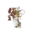
| Structure viewer | Molecule: MolmilJmol/JSmol |
|---|
- Downloads & links
-Download
| PDBx/mmCIF format | 3btf.cif.gz | 69.2 KB | Display | PDBx/mmCIF format |
|---|---|---|---|---|
| PDB format | pdb3btf.ent.gz | 49.5 KB | Display | PDB format |
| PDBx/mmJSON format | 3btf.json.gz | Tree view | PDBx/mmJSON format | |
| Others |  Other downloads Other downloads |
-Validation report
| Arichive directory | https://data.pdbj.org/pub/pdb/validation_reports/bt/3btfftp://data.pdbj.org/pub/pdb/validation_reports/bt/3btf | HTTPS FTP |
|---|
-Related structure data
| Related structure data |  3btdC  3bteC  3btgC  3bthC 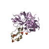 3btkC  3btmC  3btqC  3bttC  3btwC 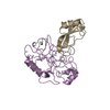 2ptcS S: Starting model for refinement C: citing same article ( |
|---|---|
| Similar structure data |


-Links
 PDBj
PDBj


- Assembly
| Deposited unit | 
| ||||||||
|---|---|---|---|---|---|---|---|---|---|
| 1 |
| ||||||||
| 2 | 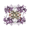
| ||||||||
| 3 | 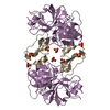
| ||||||||
| Unit cell |
|
-Components
| #1: Protein | Mass: 23324.287 Da / Num. of mol.: 1 / Source method: isolated from a natural source / Source: (natural) | ||||||
|---|---|---|---|---|---|---|---|
| #2: Protein | Mass: 6527.524 Da / Num. of mol.: 1 / Mutation: YES Source method: isolated from a genetically manipulated source Source: (gene. exp.)  | ||||||
| #3: Chemical | ChemComp-CA /   Mass: 40.078 Da / Num. of mol.: 1 / Source method: obtained synthetically / Formula: Ca Mass: 40.078 Da / Num. of mol.: 1 / Source method: obtained synthetically / Formula: Ca | ||||||
| #4: Chemical | ChemComp-SO4 /   Mass: 96.063 Da / Num. of mol.: 4 / Source method: obtained synthetically / Formula: SO4 Mass: 96.063 Da / Num. of mol.: 4 / Source method: obtained synthetically / Formula: SO4#5: Water | ChemComp-HOH / |  Mass: 18.015 Da / Num. of mol.: 131 / Source method: isolated from a natural source / Formula: H2O Mass: 18.015 Da / Num. of mol.: 131 / Source method: isolated from a natural source / Formula: H2OCompound details | P1 RESIDUE OF THE INHIBITOR IS MUTATED TO PHE | Has protein modification | Y | |
-Experimental details
-Experiment
| Experiment | Method: X-RAY DIFFRACTION / Number of used crystals: 1 |
|---|
- Sample preparation
| Crystal | Density Matthews: 3.32 Å3/Da / Density % sol: 55 % | ||||||||||||||||||||
|---|---|---|---|---|---|---|---|---|---|---|---|---|---|---|---|---|---|---|---|---|---|
| Crystal grow | pH: 7.5 / Details: 0.1 M HEPES PH 7.5 48% AMMONIUM SULPHATE | ||||||||||||||||||||
| Crystal grow | *PLUS Temperature: 37 ℃ / Method: vapor diffusion, hanging drop | ||||||||||||||||||||
| Components of the solutions | *PLUS
|
-Data collection
| Diffraction | Mean temperature: 293 K |
|---|---|
| Diffraction source | Source: SYNCHROTRON / Site: ESRF  / Beamline: BM1A / Wavelength: 0.873 / Beamline: BM1A / Wavelength: 0.873 |
| Detector | Type: MARRESEARCH / Detector: IMAGE PLATE / Date: Apr 1, 1998 |
| Radiation | Protocol: SINGLE WAVELENGTH / Monochromatic (M) / Laue (L): M / Scattering type: x-ray |
| Radiation wavelength | Wavelength: 0.873 Å / Relative weight: 1 |
| Reflection | Resolution: 1.8→15 Å / Num. obs: 36655 / % possible obs: 98.1 % / Redundancy: 4 % / Biso Wilson estimate: 20 Å2 / Rmerge(I) obs: 0.059 / Rsym value: 0.059 / Net I/σ(I): 8.4 |
| Reflection shell | Resolution: 1.8→1.9 Å / Redundancy: 4 % / Rmerge(I) obs: 0.326 / Mean I/σ(I) obs: 2.2 / Rsym value: 0.326 / % possible all: 98.8 |
| Reflection shell | *PLUS % possible obs: 98.8 % |
- Processing
| Software |
| ||||||||||||||||||||||||||||||||||||||||||||||||||||||||||||
|---|---|---|---|---|---|---|---|---|---|---|---|---|---|---|---|---|---|---|---|---|---|---|---|---|---|---|---|---|---|---|---|---|---|---|---|---|---|---|---|---|---|---|---|---|---|---|---|---|---|---|---|---|---|---|---|---|---|---|---|---|---|
| Refinement | Method to determine structure: MOLECULAR REPLACEMENT Starting model: 2PTC Resolution: 1.8→8 Å / Cross valid method: THROUGHOUT / σ(F): 1 Details: ENERGY TERMS OF THE INHIBITOR SCISSILE PEPTIDE BOND WERE SET TO ZERO DURING REFINEMENT
| ||||||||||||||||||||||||||||||||||||||||||||||||||||||||||||
| Displacement parameters | Biso mean: 21.8 Å2 | ||||||||||||||||||||||||||||||||||||||||||||||||||||||||||||
| Refinement step | Cycle: LAST / Resolution: 1.8→8 Å
| ||||||||||||||||||||||||||||||||||||||||||||||||||||||||||||
| Refine LS restraints |
| ||||||||||||||||||||||||||||||||||||||||||||||||||||||||||||
| LS refinement shell | Resolution: 1.8→1.9 Å / Total num. of bins used: 8 /
| ||||||||||||||||||||||||||||||||||||||||||||||||||||||||||||
| Xplor file |
|
