 Movie
Movie Controller

[English] 日本語
 Yorodumi
- PDB-1c1q: RECRUITING ZINC TO MEDIATE POTENT, SPECIFIC INHIBITION OF SERINE ... -
+ Open data

- Basic information
| Entry | Database: PDB / ID: 1c1q | ||||||
|---|---|---|---|---|---|---|---|
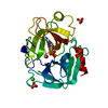
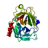
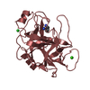
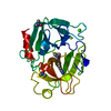
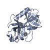
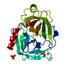
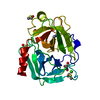
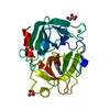
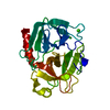
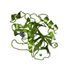
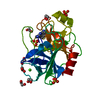
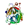
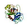
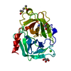
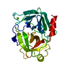
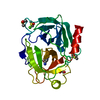
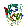
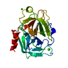
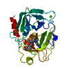
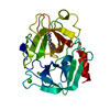
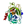
| Title | RECRUITING ZINC TO MEDIATE POTENT, SPECIFIC INHIBITION OF SERINE PROTEASES | ||||||
 Components Components | TRYPSIN | ||||||
 Keywords Keywords | HYDROLASE/HYDROLASE INHIBITOR / ZN(II)-MEDIATED SERINE PROTEASE INHIBITORS / PH DEPENDENCE / ZN(II) AFFINITY STUCTURE-BASED DRUG DESIGN / SERINE PROTEASE SERINE PROTEASE/INHIBITOR / HYDROLASE-HYDROLASE INHIBITOR COMPLEX | ||||||
| Function / homology |  Function and homology information Function and homology informationtrypsin / serpin family protein binding / serine protease inhibitor complex / digestion / endopeptidase activity / serine-type endopeptidase activity / proteolysis / : / metal ion binding Similarity search - Function | ||||||
| Biological species |  | ||||||
| Method |  X-RAY DIFFRACTION / DIFFERENCE FOURIER PLUS REFINEMENT / Resolution: 1.37 Å X-RAY DIFFRACTION / DIFFERENCE FOURIER PLUS REFINEMENT / Resolution: 1.37 Å | ||||||
 Authors Authors | Katz, B.A. / Luong, C. | ||||||
 Citation Citation | Journal: Nature / Year: 1998 Title: Design of potent selective zinc-mediated serine protease inhibitors. Authors: Katz, B.A. / Clark, J.M. / Finer-Moore, J.S. / Jenkins, T.E. / Johnson, C.R. / Ross, M.J. / Luong, C. / Moore, W.R. / Stroud, R.M. | ||||||
| History |
|
- Structure visualization
| Structure viewer | Molecule: MolmilJmol/JSmol |
|---|
- Downloads & links
-Download
| PDBx/mmCIF format | 1c1q.cif.gz | 107.5 KB | Display | PDBx/mmCIF format |
|---|---|---|---|---|
| PDB format | pdb1c1q.ent.gz | 85.3 KB | Display | PDB format |
| PDBx/mmJSON format | 1c1q.json.gz | Tree view | PDBx/mmJSON format | |
| Others |  Other downloads Other downloads |
-Validation report
| Arichive directory | https://data.pdbj.org/pub/pdb/validation_reports/c1/1c1qftp://data.pdbj.org/pub/pdb/validation_reports/c1/1c1q | HTTPS FTP |
|---|
-Related structure data
| Related structure data | 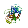 1c1nC 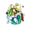 1c1oC 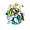 1c1pC 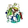 1c1rC 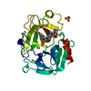 1c1tC 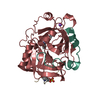 1c1uC 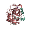 1c1vC 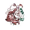 1c1wC 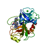 1c2dC 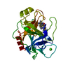 1c2eC 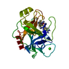 1c2fC  1c2gC 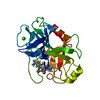 1c2hC 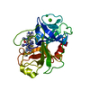 1c2iC 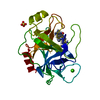 1c2jC 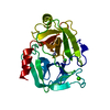 1c2kC 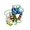 1c2lC  1c2mC 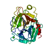 1xufC 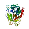 1xugC 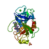 1xuhC 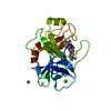 1xuiC 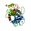 1xujC 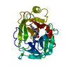 1xukC C: citing same article ( |
|---|---|
| Similar structure data |
-Links
 PDBj
PDBj

- Assembly
| Deposited unit | 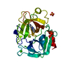
| ||||||||
|---|---|---|---|---|---|---|---|---|---|
| 1 |
| ||||||||
| Unit cell |
|
-Components
-Protein , 1 types, 1 molecules A
| #1: Protein | Mass: 23324.287 Da / Num. of mol.: 1 / Source method: isolated from a natural source Details: COMPLEXED WITH (5-AMIDINO-2-BENZIMIDAZOLYL)(2-BENZIMIDAZOLYL)METHANE Source: (natural) |
|---|
-Non-polymers , 5 types, 219 molecules 


| #2: Chemical | ChemComp-CA /  Mass: 40.078 Da / Num. of mol.: 1 / Source method: obtained synthetically / Formula: Ca Mass: 40.078 Da / Num. of mol.: 1 / Source method: obtained synthetically / Formula: Ca |
|---|---|
| #3: Chemical | ChemComp-MG /  Mass: 24.305 Da / Num. of mol.: 1 / Source method: obtained synthetically / Formula: Mg Mass: 24.305 Da / Num. of mol.: 1 / Source method: obtained synthetically / Formula: Mg |
| #4: Chemical | ChemComp-SO4 /  Mass: 96.063 Da / Num. of mol.: 1 / Source method: obtained synthetically / Formula: SO4 Mass: 96.063 Da / Num. of mol.: 1 / Source method: obtained synthetically / Formula: SO4 |
| #5: Chemical | ChemComp-BAI / ( Mass: 290.323 Da / Num. of mol.: 1 / Source method: obtained synthetically / Formula: C16H14N6 Mass: 290.323 Da / Num. of mol.: 1 / Source method: obtained synthetically / Formula: C16H14N6 |
| #6: Water | ChemComp-HOH / Mass: 18.015 Da / Num. of mol.: 215 / Source method: isolated from a natural source / Formula: H2O |
-Details
| Compound details | HIS40 AND HIS91 ARE MONOPROTON| Has protein modification | Y | |
|---|
-Experimental details
-Experiment
| Experiment | Method: X-RAY DIFFRACTION / Number of used crystals: 1 |
|---|
- Sample preparation
| Crystal | Density Matthews: 2.03 Å3/Da / Density % sol: 18 % |
|---|---|
| Crystal grow | pH: 8.21 Details: TRYPSIN-BENZAMIDINE, P3(1) 2 1 WERE GROWN BY VAPOR DIFFUSION, AS DESCRIBED FOR P2(1) 2(1) 2(1) (LARGE CELL) (MANGEL, ET AL., BIOCHEMISTRY 29, 8351-8357, 1990) THE CRYSTAL WAS SOAKED IN A ...Details: TRYPSIN-BENZAMIDINE, P3(1) 2 1 WERE GROWN BY VAPOR DIFFUSION, AS DESCRIBED FOR P2(1) 2(1) 2(1) (LARGE CELL) (MANGEL, ET AL., BIOCHEMISTRY 29, 8351-8357, 1990) THE CRYSTAL WAS SOAKED IN A SOLUTION OF 0.10 M TRIS, 2.02 M MGSO4 . 7 H2O, PH 8.21, 2.0 % DMSO, SATURATED IN HEMI-BABIM OVER A PERIOD OF SEVERAL DAYS WITH SEVERAL REPLACEMENTS OF THE SOAKING SOLUTION. |
| Crystal grow | *PLUS Method: unknown |
-Data collection
| Diffraction | Mean temperature: 298 K |
|---|---|
| Diffraction source | Source: ROTATING ANODE / Wavelength: 1.5418 |
| Detector | Type: RIGAKU RAXIS IV++ / Detector: IMAGE PLATE / Date: Jun 26, 1999 / Details: MSC MIRRORS |
| Radiation | Protocol: SINGLE WAVELENGTH / Monochromatic (M) / Laue (L): M / Scattering type: x-ray |
| Radiation wavelength | Wavelength: 1.5418 Å / Relative weight: 1 |
| Reflection | Resolution: 1.32→43.61 Å / Num. obs: 34963 / % possible obs: 82.7 % / Observed criterion σ(I): 0.8 / Redundancy: 3.4 % / Rmerge(I) obs: 0.068 / Net I/σ(I): 10.8 |
| Reflection shell | Resolution: 1.37→1.43 Å / Rmerge(I) obs: 0.268 / Mean I/σ(I) obs: 1.8 / % possible all: 35.5 |
- Processing
| Software |
| ||||||||||||||||||||||||||||||||||||||||||||||||||||||||||||
|---|---|---|---|---|---|---|---|---|---|---|---|---|---|---|---|---|---|---|---|---|---|---|---|---|---|---|---|---|---|---|---|---|---|---|---|---|---|---|---|---|---|---|---|---|---|---|---|---|---|---|---|---|---|---|---|---|---|---|---|---|---|
| Refinement | Method to determine structure: DIFFERENCE FOURIER PLUS REFINEMENT Resolution: 1.37→7.5 Å / Cross valid method: X-PLOR / σ(F): 1.8 Details: BULK SOLVENT TERMS INCLUDED IN FOB FILE CREATED WITH STANDARD X-PLOR SCRIPT.
| ||||||||||||||||||||||||||||||||||||||||||||||||||||||||||||
| Refinement step | Cycle: LAST / Resolution: 1.37→7.5 Å
| ||||||||||||||||||||||||||||||||||||||||||||||||||||||||||||
| Refine LS restraints |
| ||||||||||||||||||||||||||||||||||||||||||||||||||||||||||||
| LS refinement shell | Resolution: 1.37→1.43 Å / Total num. of bins used: 8
| ||||||||||||||||||||||||||||||||||||||||||||||||||||||||||||
| Xplor file |
|
