 Movie
Movie Controller

[English] 日本語
 Yorodumi
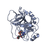
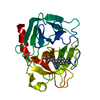
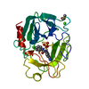
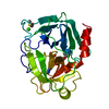
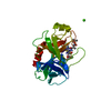
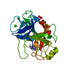
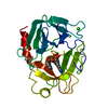
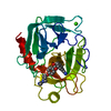
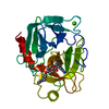
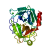
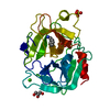
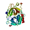
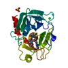
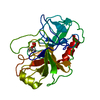
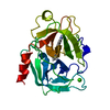
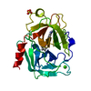
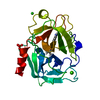
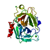
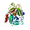
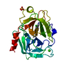
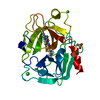
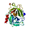
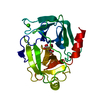
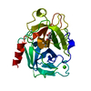
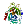
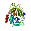
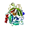
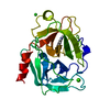
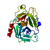
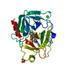
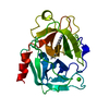
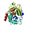
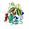
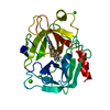
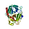
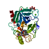
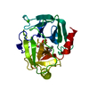
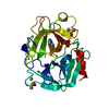
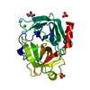
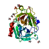
- PDB-1utn: Trypsin specificity as elucidated by LIE calculations, X-ray stru... -
+ Open data

- Basic information
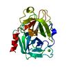
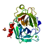
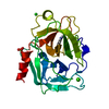
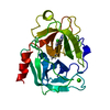
| Entry | Database: PDB / ID: 1utn | ||||||
|---|---|---|---|---|---|---|---|
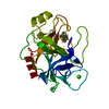
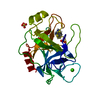
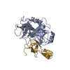
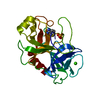
| Title | Trypsin specificity as elucidated by LIE calculations, X-ray structures and association constant measurements | ||||||
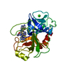
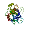
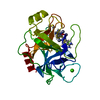
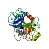
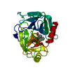
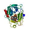
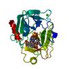
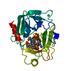
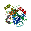
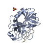
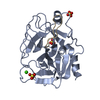
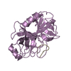
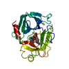
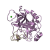
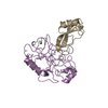
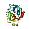
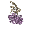
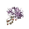
 Components Components | TRYPSINOGEN | ||||||
 Keywords Keywords | HYDROLASE / TRYPSIN / INHIBITOR SPECIFICITY / ELECTROSTATIC INTERACTIONS / COLD-ADAPTATION / MOLECULAR DYNAMICS / BINDING FREE ENERGY HYDROLASE | ||||||
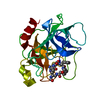
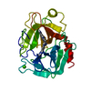
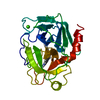
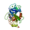
| Function / homology |  Function and homology information Function and homology informationtrypsin / serpin family protein binding / serine protease inhibitor complex / digestion / endopeptidase activity / serine-type endopeptidase activity / proteolysis / : / metal ion binding Similarity search - Function | ||||||
| Biological species |  | ||||||
| Method |  X-RAY DIFFRACTION / SYNCHROTRON / OTHER / Resolution: 1.15 Å X-RAY DIFFRACTION / SYNCHROTRON / OTHER / Resolution: 1.15 Å | ||||||
 Authors Authors | Leiros, H.-K.S. / Brandsdal, B.O. / Andersen, O.A. / Os, V. / Leiros, I. / Helland, R. / Otlewski, J. / Willassen, N.P. / Smalas, A.O. | ||||||
 Citation Citation | Journal: Protein Sci. / Year: 2004 Title: Trypsin Specificity as Elucidated by Lie Calculations, X-Ray Structures, and Association Constant Measurements Authors: Leiros, H.-K.S. / Brandsdal, B.O. / Andersen, O.A. / Os, V. / Leiros, I. / Helland, R. / Otlewski, J. / Willassen, N.P. / Smalas, A.O. | ||||||
| History |
| ||||||
| Remark 700 | SHEET DETERMINATION METHOD: DSSP THE SHEETS PRESENTED AS "AB" IN EACH CHAIN ON SHEET RECORDS BELOW ... SHEET DETERMINATION METHOD: DSSP THE SHEETS PRESENTED AS "AB" IN EACH CHAIN ON SHEET RECORDS BELOW IS ACTUALLY AN 6-STRANDED BARREL THIS IS REPRESENTED BY A 7-STRANDED SHEET IN WHICH THE FIRST AND LAST STRANDS ARE IDENTICAL. |
- Structure visualization
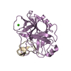
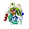
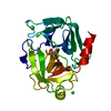
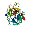
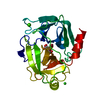
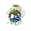
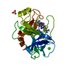
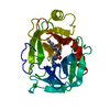
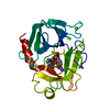
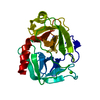
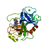
| Structure viewer | Molecule: MolmilJmol/JSmol |
|---|
- Downloads & links
-Download
| PDBx/mmCIF format | 1utn.cif.gz | 151.6 KB | Display | PDBx/mmCIF format |
|---|---|---|---|---|
| PDB format | pdb1utn.ent.gz | 122.2 KB | Display | PDB format |
| PDBx/mmJSON format | 1utn.json.gz | Tree view | PDBx/mmJSON format | |
| Others |  Other downloads Other downloads |
-Validation report
| Arichive directory | https://data.pdbj.org/pub/pdb/validation_reports/ut/1utnftp://data.pdbj.org/pub/pdb/validation_reports/ut/1utn | HTTPS FTP |
|---|
-Related structure data
| Related structure data | 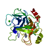 1utjC 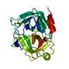 1utkC 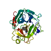 1utlC 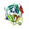 1utmC 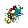 1utoC 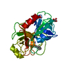 1utpC  1utqC C: citing same article ( |
|---|---|
| Similar structure data |


-Links
 PDBj
PDBj

- Assembly
| Deposited unit | 
| ||||||||
|---|---|---|---|---|---|---|---|---|---|
| 1 |
| ||||||||
| Unit cell |
|
-Components
-Protein , 1 types, 1 molecules A
| #1: Protein | Mass: 25444.717 Da / Num. of mol.: 1 / Source method: isolated from a natural source / Source: (natural) |
|---|
-Non-polymers , 6 types, 297 molecules 


| #2: Chemical |  Mass: 107.153 Da / Num. of mol.: 2 / Source method: obtained synthetically / Formula: C7H9N Mass: 107.153 Da / Num. of mol.: 2 / Source method: obtained synthetically / Formula: C7H9N#3: Chemical | ChemComp-SO4 /  Mass: 96.063 Da / Num. of mol.: 5 / Source method: obtained synthetically / Formula: SO4 Mass: 96.063 Da / Num. of mol.: 5 / Source method: obtained synthetically / Formula: SO4#4: Chemical |  Mass: 92.094 Da / Num. of mol.: 2 / Source method: obtained synthetically / Formula: C3H8O3 Mass: 92.094 Da / Num. of mol.: 2 / Source method: obtained synthetically / Formula: C3H8O3#5: Chemical | ChemComp-TRS / |  Mass: 122.143 Da / Num. of mol.: 1 / Source method: obtained synthetically / Formula: C4H12NO3 / Comment: pH buffer*YM Mass: 122.143 Da / Num. of mol.: 1 / Source method: obtained synthetically / Formula: C4H12NO3 / Comment: pH buffer*YM#6: Chemical | ChemComp-CA / |  Mass: 40.078 Da / Num. of mol.: 1 / Source method: obtained synthetically / Formula: Ca Mass: 40.078 Da / Num. of mol.: 1 / Source method: obtained synthetically / Formula: Ca#7: Water | ChemComp-HOH / | Mass: 18.015 Da / Num. of mol.: 286 / Source method: isolated from a natural source / Formula: H2O |
|---|
-Details
| Has protein modification | Y |
|---|
-Experimental details
-Experiment
| Experiment | Method: X-RAY DIFFRACTION / Number of used crystals: 1 |
|---|
- Sample preparation
| Crystal | Density Matthews: 2 Å3/Da / Density % sol: 38.49 % | ||||||||||||||||||||||||||||||||||||
|---|---|---|---|---|---|---|---|---|---|---|---|---|---|---|---|---|---|---|---|---|---|---|---|---|---|---|---|---|---|---|---|---|---|---|---|---|---|
| Crystal grow | pH: 8 / Details: pH 8.00 | ||||||||||||||||||||||||||||||||||||
| Crystal grow | *PLUS pH: 8 / Method: vapor diffusion, hanging drop | ||||||||||||||||||||||||||||||||||||
| Components of the solutions | *PLUS
|
-Data collection
| Diffraction | Mean temperature: 120 K |
|---|---|
| Diffraction source | Source: SYNCHROTRON / Site: ESRF  / Beamline: BM1A / Wavelength: 0.8 / Beamline: BM1A / Wavelength: 0.8 |
| Detector | Type: MARRESEARCH / Detector: IMAGE PLATE |
| Radiation | Protocol: SINGLE WAVELENGTH / Monochromatic (M) / Laue (L): M / Scattering type: x-ray |
| Radiation wavelength | Wavelength: 0.8 Å / Relative weight: 1 |
| Reflection | Resolution: 1.15→8 Å / Num. obs: 71444 / % possible obs: 97.3 % / Redundancy: 11.2 % / Rmerge(I) obs: 0.055 / Net I/σ(I): 7.2 |
| Reflection shell | Rmerge(I) obs: 0.356 / Mean I/σ(I) obs: 1.9 / % possible all: 91.6 |
| Reflection | *PLUS Highest resolution: 1.15 Å / Num. measured all: 801482 / Rmerge(I) obs: 0.055 |
| Reflection shell | *PLUS % possible obs: 91.6 % / Rmerge(I) obs: 0.356 / Mean I/σ(I) obs: 1.9 |
- Processing
| Software |
| |||||||||||||||||||||||||||||||||
|---|---|---|---|---|---|---|---|---|---|---|---|---|---|---|---|---|---|---|---|---|---|---|---|---|---|---|---|---|---|---|---|---|---|---|
| Refinement | Method to determine structure: OTHER / Resolution: 1.15→8 Å / Cross valid method: THROUGHOUT / σ(F): 0
| |||||||||||||||||||||||||||||||||
| Refinement step | Cycle: LAST / Resolution: 1.15→8 Å
| |||||||||||||||||||||||||||||||||
| Refine LS restraints |
| |||||||||||||||||||||||||||||||||
| Software | *PLUS Name: SHELXL / Version: 97 / Classification: refinement | |||||||||||||||||||||||||||||||||
| Refinement | *PLUS Lowest resolution: 8 Å / Rfactor Rwork: 0.1132 | |||||||||||||||||||||||||||||||||
| Solvent computation | *PLUS | |||||||||||||||||||||||||||||||||
| Displacement parameters | *PLUS |
