 Movie
Movie Controller

[English] 日本語
 Yorodumi
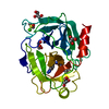
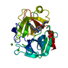
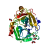
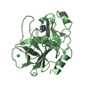
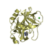
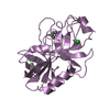
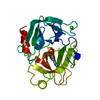
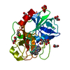
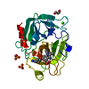
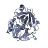
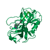
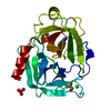
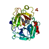
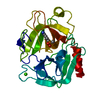
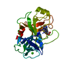
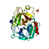
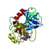
- PDB-1c5p: STRUCTURAL BASIS FOR SELECTIVITY OF A SMALL MOLECULE, S1-BINDING,... -
+ Open data

- Basic information
| Entry | Database: PDB / ID: 1c5p | |||||||||
|---|---|---|---|---|---|---|---|---|---|---|
| Title | STRUCTURAL BASIS FOR SELECTIVITY OF A SMALL MOLECULE, S1-BINDING, SUB-MICROMOLAR INHIBITOR OF UROKINASE TYPE PLASMINOGEN ACTIVATOR | |||||||||
 Components Components | PROTEIN (TRYPSIN) | |||||||||
 Keywords Keywords | HYDROLASE / selective / S1 site inhibitor / structure-based drug design / urokinase / trypsin / thrombin | |||||||||
| Function / homology |  Function and homology information Function and homology informationtrypsin / serpin family protein binding / serine protease inhibitor complex / digestion / endopeptidase activity / serine-type endopeptidase activity / proteolysis / : / metal ion binding Similarity search - Function | |||||||||
| Biological species |  | |||||||||
| Method |  X-RAY DIFFRACTION / DIFFERENCE FOURIER PLUS REFINEMENT / Resolution: 1.43 Å X-RAY DIFFRACTION / DIFFERENCE FOURIER PLUS REFINEMENT / Resolution: 1.43 Å | |||||||||
 Authors Authors | Katz, B.A. / Mackman, R. / Luong, C. / Radika, K. / Martelli, A. / Sprengeler, P.A. / Wang, J. / Chan, H. / Wong, L. | |||||||||
 Citation Citation | Journal: Chem.Biol. / Year: 2000 Title: Structural basis for selectivity of a small molecule, S1-binding, submicromolar inhibitor of urokinase-type plasminogen activator. Authors: Katz, B.A. / Mackman, R. / Luong, C. / Radika, K. / Martelli, A. / Sprengeler, P.A. / Wang, J. / Chan, H. / Wong, L. | |||||||||
| History |
|
- Structure visualization
| Structure viewer | Molecule: MolmilJmol/JSmol |
|---|
- Downloads & links
-Download
| PDBx/mmCIF format | 1c5p.cif.gz | 106.8 KB | Display | PDBx/mmCIF format |
|---|---|---|---|---|
| PDB format | pdb1c5p.ent.gz | 84.4 KB | Display | PDB format |
| PDBx/mmJSON format | 1c5p.json.gz | Tree view | PDBx/mmJSON format | |
| Others |  Other downloads Other downloads |
-Validation report
| Arichive directory | https://data.pdbj.org/pub/pdb/validation_reports/c5/1c5pftp://data.pdbj.org/pub/pdb/validation_reports/c5/1c5p | HTTPS FTP |
|---|
-Related structure data
| Related structure data | 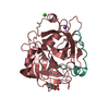 1c5lC 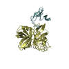 1c5mC  1c5nC 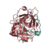 1c5oC 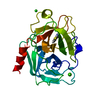 1c5qC 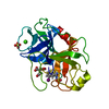 1c5rC 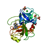 1c5sC 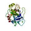 1c5tC 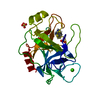 1c5uC  1c5vC 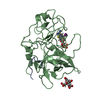 1c5wC 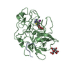 1c5xC 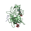 1c5yC 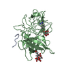 1c5zC C: citing same article ( |
|---|---|
| Similar structure data |

-Links
 PDBj
PDBj


- Assembly
| Deposited unit | 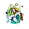
| ||||||||
|---|---|---|---|---|---|---|---|---|---|
| 1 |
| ||||||||
| Unit cell |
| ||||||||
| Components on special symmetry positions |
|
-Components
-Protein , 1 types, 1 molecules A
| #1: Protein | Mass: 23324.287 Da / Num. of mol.: 1 / Source method: isolated from a natural source / Source: (natural) |
|---|
-Non-polymers , 5 types, 223 molecules 


| #2: Chemical | ChemComp-CA /  Mass: 40.078 Da / Num. of mol.: 1 / Source method: obtained synthetically / Formula: Ca Mass: 40.078 Da / Num. of mol.: 1 / Source method: obtained synthetically / Formula: Ca | ||||
|---|---|---|---|---|---|
| #3: Chemical | ChemComp-MG /  Mass: 24.305 Da / Num. of mol.: 1 / Source method: obtained synthetically / Formula: Mg Mass: 24.305 Da / Num. of mol.: 1 / Source method: obtained synthetically / Formula: Mg | ||||
| #4: Chemical |  Mass: 96.063 Da / Num. of mol.: 2 / Source method: obtained synthetically / Formula: SO4 Mass: 96.063 Da / Num. of mol.: 2 / Source method: obtained synthetically / Formula: SO4#5: Chemical | ChemComp-BEN / |  Mass: 120.152 Da / Num. of mol.: 1 / Source method: obtained synthetically / Formula: C7H8N2 Mass: 120.152 Da / Num. of mol.: 1 / Source method: obtained synthetically / Formula: C7H8N2#6: Water | ChemComp-HOH / | Mass: 18.015 Da / Num. of mol.: 218 / Source method: isolated from a natural source / Formula: H2O |
-Details
| Has protein modification | Y |
|---|
-Experimental details
-Experiment
| Experiment | Method: X-RAY DIFFRACTION / Number of used crystals: 1 |
|---|
- Sample preparation
| Crystal | Density Matthews: 2.03 Å3/Da / Density % sol: 18 % | ||||||||||||||||||||||||||||||
|---|---|---|---|---|---|---|---|---|---|---|---|---|---|---|---|---|---|---|---|---|---|---|---|---|---|---|---|---|---|---|---|
| Crystal grow | Method: vapor diffusion / pH: 7.5 Details: trypsin-benzamidine, P2(1)2(1)2(1) were grown by vapor diffusion, as described for P2(1) 2(1) 2(1) (large cell) (Mangel, et al., Biochemistry 29, 8351-8357, 1990) The crystal was soaked in a ...Details: trypsin-benzamidine, P2(1)2(1)2(1) were grown by vapor diffusion, as described for P2(1) 2(1) 2(1) (large cell) (Mangel, et al., Biochemistry 29, 8351-8357, 1990) The crystal was soaked in a solution of 85 % saturated MgSO4 . 7 H2O, 1 mM CaCl2, 2.0 % DMSO, 100 mM Tris, pH 7.50., VAPOR DIFFUSION | ||||||||||||||||||||||||||||||
| Crystal grow | *PLUS pH: 7.5 / Method: batch method / Details: Katz, B.A., (1999) J. Mol. Biol., 292, 669. | ||||||||||||||||||||||||||||||
| Components of the solutions | *PLUS
|
-Data collection
| Diffraction | Mean temperature: 298 K |
|---|---|
| Diffraction source | Source: ROTATING ANODE / Wavelength: 1.5418 |
| Detector | Type: RIGAKU RAXIS IV++ / Detector: IMAGE PLATE / Date: May 23, 1997 / Details: MSC MIRRORS |
| Radiation | Protocol: SINGLE WAVELENGTH / Monochromatic (M) / Laue (L): M / Scattering type: x-ray |
| Radiation wavelength | Wavelength: 1.5418 Å / Relative weight: 1 |
| Reflection | Resolution: 1.21→31.93 Å / Num. all: 426850 / % possible obs: 88 % / Observed criterion σ(I): 1 / Redundancy: 8.1 % / Rmerge(I) obs: 0.09 |
| Reflection shell | Resolution: 1.21→1.26 Å / % possible all: 42.6 |
| Reflection | *PLUS Rmerge(I) obs: 0.09 |
- Processing
| Software |
| |||||||||||||||||||||||||||
|---|---|---|---|---|---|---|---|---|---|---|---|---|---|---|---|---|---|---|---|---|---|---|---|---|---|---|---|---|
| Refinement | Method to determine structure: DIFFERENCE FOURIER PLUS REFINEMENT Resolution: 1.43→7.5 Å / Cross valid method: X-PLOR / σ(F): 1.7 Details: Bulk solvent terms included in Fob file created with standard X-PLOR script. Residues simultaneously refined in two or more conformations are: Gln50, Val53, Ser61, Leu67, Lys87, Met104, ...Details: Bulk solvent terms included in Fob file created with standard X-PLOR script. Residues simultaneously refined in two or more conformations are: Gln50, Val53, Ser61, Leu67, Lys87, Met104, Ser110, Ser170, Ser236. Disordered waters are: HOH344 which is close to HOH347; HOH349 which is close to HOH601 which is close to HOH382; HOH365 which is close to HOH366; HOH702 which is close to HOH704; HOH789 which is close to a symmetry-related equivalent of itself. HOH371 WHICH IS CLOSE TO A SYMMETRY_RELATED EQUIVALENT OF ITSELF. HOH859 WHICH IS CLOSE TO HOH861; HOH296 WHICH IS CLOSE TO A SYMMETRY_RELATED EQUIVALENT OF ITSELF; HIS91 is MONOPROTONATED ON THE EPSILON NITROGEN. HIS 40 and HIS57 are DIPROTONATED.
| |||||||||||||||||||||||||||
| Refinement step | Cycle: LAST / Resolution: 1.43→7.5 Å
| |||||||||||||||||||||||||||
| Refine LS restraints |
| |||||||||||||||||||||||||||
| LS refinement shell | Resolution: 1.43→1.49 Å / Total num. of bins used: 8
| |||||||||||||||||||||||||||
| Xplor file |
| |||||||||||||||||||||||||||
| Software | *PLUS Name: X-PLOR / Version: 3.1 / Classification: refinement | |||||||||||||||||||||||||||
| Refinement | *PLUS Highest resolution: 1.21 Å / Lowest resolution: 7.5 Å / σ(F): 1.7 / % reflection Rfree: 10 % / Rfactor obs: 0.194 | |||||||||||||||||||||||||||
| Solvent computation | *PLUS | |||||||||||||||||||||||||||
| Displacement parameters | *PLUS | |||||||||||||||||||||||||||
| Refine LS restraints | *PLUS
| |||||||||||||||||||||||||||
| LS refinement shell | *PLUS Rfactor Rfree: 0.382 / Rfactor Rwork: 0.375 |
