[English] 日本語
 Yorodumi
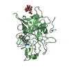
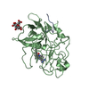
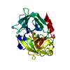
- PDB-1c5w: STRUCTURAL BASIS FOR SELECTIVITY OF A SMALL MOLECULE, S1-BINDING,... -
+ Open data
- Basic information
| Entry | Database: PDB / ID: 1c5w | |||||||||
|---|---|---|---|---|---|---|---|---|---|---|
| Title | STRUCTURAL BASIS FOR SELECTIVITY OF A SMALL MOLECULE, S1-BINDING, SUB-MICROMOLAR INHIBITOR OF UROKINASE TYPE PLASMINOGEN ACTIVATOR | |||||||||
 Components Components | (PROTEIN (UROKINASE-TYPE PLASMINOGEN ACTIVATOR)) x 2 | |||||||||
 Keywords Keywords | BLOOD CLOTTING / selective / S1 site inhibitor / structure-based drug design / urokinase / trypsin / thrombin | |||||||||
| Function / homology |  Function and homology information Function and homology informationu-plasminogen activator / regulation of smooth muscle cell-matrix adhesion / regulation of integrin-mediated signaling pathway / urokinase plasminogen activator signaling pathway / regulation of plasminogen activation / regulation of fibrinolysis / protein complex involved in cell-matrix adhesion / regulation of wound healing / negative regulation of plasminogen activation / serine-type endopeptidase complex ...u-plasminogen activator / regulation of smooth muscle cell-matrix adhesion / regulation of integrin-mediated signaling pathway / urokinase plasminogen activator signaling pathway / regulation of plasminogen activation / regulation of fibrinolysis / protein complex involved in cell-matrix adhesion / regulation of wound healing / negative regulation of plasminogen activation / serine-type endopeptidase complex / regulation of smooth muscle cell migration / Dissolution of Fibrin Clot / smooth muscle cell migration / plasminogen activation / regulation of cell adhesion mediated by integrin / tertiary granule membrane / negative regulation of fibrinolysis / regulation of cell adhesion / positive regulation of epidermal growth factor receptor signaling pathway / serine protease inhibitor complex / specific granule membrane / fibrinolysis / chemotaxis / blood coagulation / regulation of cell population proliferation / response to hypoxia / positive regulation of cell migration / receptor ligand activity / serine-type endopeptidase activity / external side of plasma membrane / focal adhesion / Neutrophil degranulation / cell surface / signal transduction / proteolysis / extracellular space / extracellular exosome / extracellular region / plasma membrane Similarity search - Function | |||||||||
| Biological species |  Homo sapiens (human) Homo sapiens (human) | |||||||||
| Method |  X-RAY DIFFRACTION / DIFFERENCE FOURIER PLUS REFINEMENT / Resolution: 1.94 Å X-RAY DIFFRACTION / DIFFERENCE FOURIER PLUS REFINEMENT / Resolution: 1.94 Å | |||||||||
 Authors Authors | Katz, B.A. / Mackman, R. / Luong, C. / Radika, K. / Martelli, A. / Sprengeler, P.A. / Wang, J. / Chan, H. / Wong, L. | |||||||||
 Citation Citation |  Journal: Chem.Biol. / Year: 2000 Journal: Chem.Biol. / Year: 2000Title: Structural basis for selectivity of a small molecule, S1-binding, submicromolar inhibitor of urokinase-type plasminogen activator. Authors: Katz, B.A. / Mackman, R. / Luong, C. / Radika, K. / Martelli, A. / Sprengeler, P.A. / Wang, J. / Chan, H. / Wong, L. | |||||||||
| History |
|
- Structure visualization
| Structure viewer | Molecule:  Molmil Molmil Jmol/JSmol Jmol/JSmol |
|---|
- Downloads & links
- Download
| PDBx/mmCIF format |  1c5w.cif.gz 1c5w.cif.gz | 133.8 KB | Display |  PDBx/mmCIF format PDBx/mmCIF format |
|---|---|---|---|---|
| PDB format |  pdb1c5w.ent.gz pdb1c5w.ent.gz | 106.8 KB | Display |  PDB format PDB format |
| PDBx/mmJSON format |  1c5w.json.gz 1c5w.json.gz | Tree view |  PDBx/mmJSON format PDBx/mmJSON format | |
| Others |  Other downloads Other downloads |
-Validation report
| Arichive directory |  https://data.pdbj.org/pub/pdb/validation_reports/c5/1c5w https://data.pdbj.org/pub/pdb/validation_reports/c5/1c5w ftp://data.pdbj.org/pub/pdb/validation_reports/c5/1c5w ftp://data.pdbj.org/pub/pdb/validation_reports/c5/1c5w | HTTPS FTP |
|---|
-Related structure data
| Related structure data | 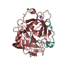 1c5lC 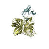 1c5mC 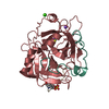 1c5nC  1c5oC  1c5pC  1c5qC  1c5rC 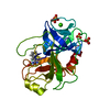 1c5sC 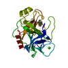 1c5tC 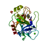 1c5uC  1c5vC 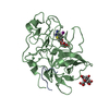 1c5xC 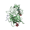 1c5yC 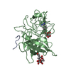 1c5zC C: citing same article ( |
|---|---|
| Similar structure data |
- Links
Links
- Assembly
| Deposited unit | 
| |||||||||||||||
|---|---|---|---|---|---|---|---|---|---|---|---|---|---|---|---|---|
| 1 |
| |||||||||||||||
| Unit cell |
| |||||||||||||||
| Components on special symmetry positions |
|
- Components
| #1: Protein/peptide | Mass: 2708.183 Da / Num. of mol.: 1 / Fragment: SHORT CHAIN Source method: isolated from a genetically manipulated source Source: (gene. exp.)  Homo sapiens (human) / Plasmid: PPIC9 / Production host: Homo sapiens (human) / Plasmid: PPIC9 / Production host:  Pichia pastoris (fungus) / Strain (production host): HISTIDINE DEFICIENT GS115 / References: UniProt: P00749, u-plasminogen activator Pichia pastoris (fungus) / Strain (production host): HISTIDINE DEFICIENT GS115 / References: UniProt: P00749, u-plasminogen activator | ||||||
|---|---|---|---|---|---|---|---|
| #2: Protein | Mass: 28435.428 Da / Num. of mol.: 1 / Fragment: CATALYTIC DOMAIN Source method: isolated from a genetically manipulated source Source: (gene. exp.)  Homo sapiens (human) / Plasmid: PPIC9 / Production host: Homo sapiens (human) / Plasmid: PPIC9 / Production host:  Pichia pastoris (fungus) / Strain (production host): HISTIDINE DEFICIENT GS115 / References: UniProt: P00749, u-plasminogen activator Pichia pastoris (fungus) / Strain (production host): HISTIDINE DEFICIENT GS115 / References: UniProt: P00749, u-plasminogen activator | ||||||
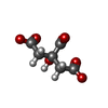
| #3: Chemical | | #4: Chemical | ChemComp-ESI / | #5: Water | ChemComp-HOH / | Has protein modification | Y | |
-Experimental details
-Experiment
| Experiment | Method:  X-RAY DIFFRACTION / Number of used crystals: 1 X-RAY DIFFRACTION / Number of used crystals: 1 |
|---|
- Sample preparation
| Crystal | Density Matthews: 2 Å3/Da / Density % sol: 25.6 % |
|---|---|
| Crystal grow | pH: 6.5 Details: LMW human uPA/A145 was concentrated to 10 mg/ml and incubated in 50 mM HEPES, 5.0 mM NaCl. pH 7.0, 1.4 mM 4-iodobenzo[b]thiophene-2-carboxamidine for 15 min on ice. The complex was ...Details: LMW human uPA/A145 was concentrated to 10 mg/ml and incubated in 50 mM HEPES, 5.0 mM NaCl. pH 7.0, 1.4 mM 4-iodobenzo[b]thiophene-2-carboxamidine for 15 min on ice. The complex was crystallized by vapor diffusion in hanging drops containing equal volumes of protein-inhibitor solution (0.28 mM uPA/A145, 1.4 mM inhibitor) and well solution (20 % 2-propanol, 20 % PEG 4K, 100 mM sodium citrate, pH 6.5) sealed over the well. |
-Data collection
| Diffraction | Mean temperature: 298 K |
|---|---|
| Diffraction source | Source:  ROTATING ANODE / Wavelength: 1.5418 ROTATING ANODE / Wavelength: 1.5418 |
| Detector | Type: RIGAKU RAXIS IV++ / Detector: IMAGE PLATE / Date: Mar 22, 1998 / Details: MSC MIRRORS |
| Radiation | Protocol: SINGLE WAVELENGTH / Monochromatic (M) / Laue (L): M / Scattering type: x-ray |
| Radiation wavelength | Wavelength: 1.5418 Å / Relative weight: 1 |
| Reflection | Resolution: 1.7→41.52 Å / Num. all: 15791 / % possible obs: 79 % / Observed criterion σ(I): 1 / Redundancy: 2.4 % / Rmerge(I) obs: 0.076 / Net I/σ(I): 6.3 |
| Reflection shell | Resolution: 1.94→2.03 Å / Rmerge(I) obs: 0.195 / Mean I/σ(I) obs: 2.3 / % possible all: 49.5 |
- Processing
| Software |
| |||||||||||||||||||||||||||
|---|---|---|---|---|---|---|---|---|---|---|---|---|---|---|---|---|---|---|---|---|---|---|---|---|---|---|---|---|
| Refinement | Method to determine structure: DIFFERENCE FOURIER PLUS REFINEMENT Starting model: PDB1LMW Resolution: 1.94→7.5 Å / Cross valid method: X-PLOR / σ(F): 2 Details: Bulk solvent terms included in Fob file created with standard X-PLOR script. Only Leu A9 to Thr_A17 are included for the A-chain. Residues prior and after these residues are not visible ...Details: Bulk solvent terms included in Fob file created with standard X-PLOR script. Only Leu A9 to Thr_A17 are included for the A-chain. Residues prior and after these residues are not visible (disordered). Residues after Glu_B245 are not visible (disordered). Residues simultaneously refined in two or more conformations are: Ile_B17, Val_B38, Met_B47, Met_B81, Glu_B86, Leu_B123, Thr_B139, Arg_B166, Gln_B192, Lys_B223, Ser_B232 No energy terms between citrate 1 and 2 are included because they appear to be hydrogen-bonded to one another via an unusually short hydrogen bond between carboxylate groups. Disordered waters are: HOH33 which is close to HOH36; HOH303 which is close to HOH306; HOH715 which is close to HOH720; HOH945 which is close to a symmetry-related equivalent of itself; HOH946 which is close to a symmetry-related equivalent of itself; HOH838 which is close to HOH853.
| |||||||||||||||||||||||||||
| Refinement step | Cycle: LAST / Resolution: 1.94→7.5 Å
| |||||||||||||||||||||||||||
| Refine LS restraints |
| |||||||||||||||||||||||||||
| LS refinement shell | Resolution: 1.03→1.94 Å / Total num. of bins used: 8
| |||||||||||||||||||||||||||
| Xplor file |
|
 Movie
Movie Controller




 PDBj
PDBj


