 Movie
Movie Controller

[English] 日本語
 Yorodumi
- PDB-1c5m: STRUCTURAL BASIS FOR SELECTIVITY OF A SMALL MOLECULE, S1-BINDING,... -
+ Open data

- Basic information
| Entry | Database: PDB / ID: 1c5m | ||||||
|---|---|---|---|---|---|---|---|
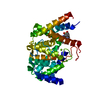
| Title | STRUCTURAL BASIS FOR SELECTIVITY OF A SMALL MOLECULE, S1-BINDING, SUB-MICROMOLAR INHIBITOR OF UROKINASE TYPE PLASMINOGEN ACTIVATOR | ||||||
 Components Components | (PROTEIN (COAGULATION FACTOR X)) x 2 | ||||||
 Keywords Keywords | BLOOD CLOTTING / selective / S1 site inhibitor / structure-based drug design / urokinase / trypsin / thrombin | ||||||
| Function / homology |  Function and homology information Function and homology informationcoagulation factor Xa / Defective factor IX causes thrombophilia / Defective cofactor function of FVIIIa variant / Defective F9 variant does not activate FX / : / positive regulation of TOR signaling / Transport of gamma-carboxylated protein precursors from the endoplasmic reticulum to the Golgi apparatus / : / Gamma-carboxylation of protein precursors / Removal of aminoterminal propeptides from gamma-carboxylated proteins ...coagulation factor Xa / Defective factor IX causes thrombophilia / Defective cofactor function of FVIIIa variant / Defective F9 variant does not activate FX / : / positive regulation of TOR signaling / Transport of gamma-carboxylated protein precursors from the endoplasmic reticulum to the Golgi apparatus / : / Gamma-carboxylation of protein precursors / Removal of aminoterminal propeptides from gamma-carboxylated proteins / : / phospholipid binding / Golgi lumen / blood coagulation / positive regulation of cell migration / endoplasmic reticulum lumen / serine-type endopeptidase activity / external side of plasma membrane / calcium ion binding / proteolysis / : / extracellular region / plasma membrane Similarity search - Function | ||||||
| Biological species |  Homo sapiens (human) Homo sapiens (human) | ||||||
| Method |  X-RAY DIFFRACTION / Resolution: 1.95 Å X-RAY DIFFRACTION / Resolution: 1.95 Å | ||||||
 Authors Authors | Katz, B.A. / Mackman, R. / Luong, C. / Radika, K. / Martelli, A. / Sprengeler, P.A. / Wang, J. / Chan, H. / Wong, L. | ||||||
 Citation Citation | Journal: Chem.Biol. / Year: 2000 Title: Structural basis for selectivity of a small molecule, S1-binding, submicromolar inhibitor of urokinase-type plasminogen activator. Authors: Katz, B.A. / Mackman, R. / Luong, C. / Radika, K. / Martelli, A. / Sprengeler, P.A. / Wang, J. / Chan, H. / Wong, L. | ||||||
| History |
|
- Structure visualization
| Structure viewer | Molecule: MolmilJmol/JSmol |
|---|
- Downloads & links
-Download
| PDBx/mmCIF format | 1c5m.cif.gz | 156.3 KB | Display | PDBx/mmCIF format |
|---|---|---|---|---|
| PDB format | pdb1c5m.ent.gz | 123.9 KB | Display | PDB format |
| PDBx/mmJSON format | 1c5m.json.gz | Tree view | PDBx/mmJSON format | |
| Others |  Other downloads Other downloads |
-Validation report
| Arichive directory | https://data.pdbj.org/pub/pdb/validation_reports/c5/1c5mftp://data.pdbj.org/pub/pdb/validation_reports/c5/1c5m | HTTPS FTP |
|---|
-Related structure data
| Related structure data |  1c5lC  1c5nC  1c5oC  1c5pC  1c5qC 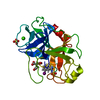 1c5rC 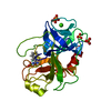 1c5sC 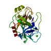 1c5tC 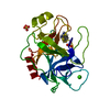 1c5uC  1c5vC  1c5wC  1c5xC  1c5yC  1c5zC  1fxaS S: Starting model for refinement C: citing same article ( |
|---|---|
| Similar structure data |


-Links
 PDBj
PDBj


- Assembly
| Deposited unit | 
| |||||||||
|---|---|---|---|---|---|---|---|---|---|---|
| 1 |
| |||||||||
| Unit cell |
| |||||||||
| Components on special symmetry positions |
|
-Components
| #1: Protein | Mass: 28649.725 Da / Num. of mol.: 1 / Fragment: HEAVY CHAIN / Source method: isolated from a natural source / Source: (natural) Homo sapiens (human) / References: UniProt: P00742, coagulation factor Xa |
|---|---|
| #2: Protein | Mass: 10533.713 Da / Num. of mol.: 1 / Fragment: LIGHT CHAIN / Source method: isolated from a natural source / Source: (natural) Homo sapiens (human) / References: UniProt: P00742, coagulation factor Xa |
| #3: Water | ChemComp-HOH /  Mass: 18.015 Da / Num. of mol.: 454 / Source method: isolated from a natural source / Formula: H2O Mass: 18.015 Da / Num. of mol.: 454 / Source method: isolated from a natural source / Formula: H2O |
| Has protein modification | Y |
-Experimental details
-Experiment
| Experiment | Method: X-RAY DIFFRACTION / Number of used crystals: 1 |
|---|
- Sample preparation
| Crystal | Density Matthews: 2.68 Å3/Da / Density % sol: 49 % | |||||||||||||||||||||||||||||||||||
|---|---|---|---|---|---|---|---|---|---|---|---|---|---|---|---|---|---|---|---|---|---|---|---|---|---|---|---|---|---|---|---|---|---|---|---|---|
| Crystal grow | Method: vapor diffusion / pH: 7.5 Details: crystallization conditions : Crystals of truncated human factor Xa were grown in hanging drops from equal volumes of protein solution (5.0 mg/ml in 20 mM Hepes, 50 mM ammonimum sulfate, pH 8. ...Details: crystallization conditions : Crystals of truncated human factor Xa were grown in hanging drops from equal volumes of protein solution (5.0 mg/ml in 20 mM Hepes, 50 mM ammonimum sulfate, pH 8.0) and well solution (25 % PEG 5K, 0.10 M Hepes, 0.2 M ammonium sulfate, pH 7.5)., VAPOR DIFFUSION | |||||||||||||||||||||||||||||||||||
| Crystal grow | *PLUS Method: vapor diffusion, hanging drop | |||||||||||||||||||||||||||||||||||
| Components of the solutions | *PLUS
|
-Data collection
| Diffraction | Mean temperature: 298 K |
|---|---|
| Diffraction source | Source: ROTATING ANODE / Wavelength: 1.5418 |
| Detector | Type: RIGAKU RAXIS IV++ / Detector: IMAGE PLATE / Date: Mar 4, 1999 / Details: MSC MIRRORS |
| Radiation | Protocol: SINGLE WAVELENGTH / Monochromatic (M) / Laue (L): M / Scattering type: x-ray |
| Radiation wavelength | Wavelength: 1.5418 Å / Relative weight: 1 |
| Reflection | Resolution: 1.64→70.86 Å / Num. all: 21969 / % possible obs: 60 % / Observed criterion σ(I): 1 / Redundancy: 1.9 % / Rmerge(I) obs: 0.072 / Net I/σ(I): 6.5 |
| Reflection shell | Resolution: 1.95→2.04 Å / Rmerge(I) obs: 0.265 / Mean I/σ(I) obs: 1.6 / % possible all: 32.2 |
- Processing
| Software |
| |||||||||||||||||||||||||||
|---|---|---|---|---|---|---|---|---|---|---|---|---|---|---|---|---|---|---|---|---|---|---|---|---|---|---|---|---|
| Refinement | Starting model: 1FXA Resolution: 1.95→7.5 Å / Cross valid method: X-PLOR / σ(F): 2 Details: Bulk solvent terms included in Fob file created with standard X-PLOR script. Residues simultaneously refined in two or more conformations are: His_F13 Disordered waters are: HOH129 which is ...Details: Bulk solvent terms included in Fob file created with standard X-PLOR script. Residues simultaneously refined in two or more conformations are: His_F13 Disordered waters are: HOH129 which is close to a symmetry-related equivalent of itself; HOH294 which is close to a symmetry-related equivalent of itself; HOH370 which is close to HOH371; HOH487 which is close to HOH490; HOH536 which is close to a symmetry-related equivalent of itself; HOH549 which is close to a symmetry-related equivalent of itself; HOH566 which is close to a symmetry-related equivalent of itself; HOH1123 which is close to a symmetry-related equivalent of itself; Waters whose occupancies are greater than unity may be due to ions more electron rich than water. Those waters with occupancies greater than unity are: HOH7, HOH8, HOH466, HOH536, HOH630, HOH671.
| |||||||||||||||||||||||||||
| Refinement step | Cycle: LAST / Resolution: 1.95→7.5 Å
| |||||||||||||||||||||||||||
| Refine LS restraints |
| |||||||||||||||||||||||||||
| LS refinement shell | Resolution: 1.95→2.04 Å / Total num. of bins used: 8
| |||||||||||||||||||||||||||
| Xplor file |
| |||||||||||||||||||||||||||
| Software | *PLUS Name: X-PLOR / Version: 3.1 / Classification: refinement | |||||||||||||||||||||||||||
| Refinement | *PLUS Lowest resolution: 7.5 Å / σ(F): 2 / % reflection Rfree: 10 % / Rfactor obs: 0.226 | |||||||||||||||||||||||||||
| Solvent computation | *PLUS | |||||||||||||||||||||||||||
| Displacement parameters | *PLUS | |||||||||||||||||||||||||||
| Refine LS restraints | *PLUS
| |||||||||||||||||||||||||||
| LS refinement shell | *PLUS Rfactor Rfree: 0.502 / % reflection Rfree: 10 % / Rfactor Rwork: 0.399 |
