 Movie
Movie Controller

[English] 日本語
 Yorodumi
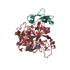
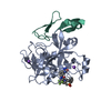
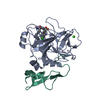
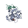
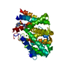
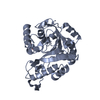
- PDB-1hcg: STRUCTURE OF HUMAN DES(1-45) FACTOR XA AT 2.2 ANGSTROMS RESOLUTION -
+ Open data

- Basic information
| Entry | Database: PDB / ID: 1hcg | ||||||
|---|---|---|---|---|---|---|---|
| Title | STRUCTURE OF HUMAN DES(1-45) FACTOR XA AT 2.2 ANGSTROMS RESOLUTION | ||||||
 Components Components | (BLOOD COAGULATION FACTOR XA) x 2 | ||||||
 Keywords Keywords | COAGULATION FACTOR | ||||||
| Function / homology |  Function and homology information Function and homology informationcoagulation factor Xa / Defective factor IX causes thrombophilia / Defective cofactor function of FVIIIa variant / Defective F9 variant does not activate FX / : / positive regulation of TOR signaling / Transport of gamma-carboxylated protein precursors from the endoplasmic reticulum to the Golgi apparatus / : / Gamma-carboxylation of protein precursors / Removal of aminoterminal propeptides from gamma-carboxylated proteins ...coagulation factor Xa / Defective factor IX causes thrombophilia / Defective cofactor function of FVIIIa variant / Defective F9 variant does not activate FX / : / positive regulation of TOR signaling / Transport of gamma-carboxylated protein precursors from the endoplasmic reticulum to the Golgi apparatus / : / Gamma-carboxylation of protein precursors / Removal of aminoterminal propeptides from gamma-carboxylated proteins / : / phospholipid binding / Golgi lumen / blood coagulation / positive regulation of cell migration / endoplasmic reticulum lumen / serine-type endopeptidase activity / external side of plasma membrane / calcium ion binding / proteolysis / : / extracellular region / plasma membrane Similarity search - Function | ||||||
| Biological species |  Homo sapiens (human) Homo sapiens (human) | ||||||
| Method |  X-RAY DIFFRACTION / Resolution: 2.2 Å X-RAY DIFFRACTION / Resolution: 2.2 Å | ||||||
 Authors Authors | Tulinsky, A. / Padmanabhan, K. | ||||||
 Citation Citation | Journal: J.Mol.Biol. / Year: 1993 Title: Structure of human des(1-45) factor Xa at 2.2 A resolution. Authors: Padmanabhan, K. / Padmanabhan, K.P. / Tulinsky, A. / Park, C.H. / Bode, W. / Huber, R. / Blankenship, D.T. / Cardin, A.D. / Kisiel, W. #1: Journal: Biochemistry / Year: 1992Title: Three-Dimensional Structure of the Apo Form of the N-Terminal Egf-Like Module of Blood Coagulation Factor X as Determined by NMR Spectroscopy and Simulated Folding Authors: Ullner, M. / Selander, M. / Persson, E. / Stenflo, J. / Drakenberg, T. / Teleman, O. #2: Journal: J.Mol.Biol. / Year: 1991Title: Structure of the Hirugen and Hirulog 1 Complexes of Alpha-Thrombin Authors: Skrzypczak-Jankun, E. / Carperos, V.E. / Ravichandran, K.G. / Tulinsky, A. / Westbrook, M. / Maraganore, J.M. #3: Journal: J.Mol.Biol. / Year: 1981Title: Comparative Model-Building of the Mammalian Serine Proteases Authors: Greer, J. | ||||||
| History |
|
- Structure visualization
| Structure viewer | Molecule: MolmilJmol/JSmol |
|---|
- Downloads & links
-Download
| PDBx/mmCIF format | 1hcg.cif.gz | 75.3 KB | Display | PDBx/mmCIF format |
|---|---|---|---|---|
| PDB format | pdb1hcg.ent.gz | 55.7 KB | Display | PDB format |
| PDBx/mmJSON format | 1hcg.json.gz | Tree view | PDBx/mmJSON format | |
| Others |  Other downloads Other downloads |
-Validation report
| Arichive directory | https://data.pdbj.org/pub/pdb/validation_reports/hc/1hcgftp://data.pdbj.org/pub/pdb/validation_reports/hc/1hcg | HTTPS FTP |
|---|
-Related structure data
| Similar structure data |
|---|


-Links
 PDBj
PDBj


- Assembly
| Deposited unit | 
| ||||||||
|---|---|---|---|---|---|---|---|---|---|
| 1 |
| ||||||||
| Unit cell |
| ||||||||
| Atom site foot note | 1: RESIDUES LEU A 247 AND GLN B 10 HAVE RAMACHANDRAN ANGLES OUTSIDE THE ALLOWED REGION. THESE RESIDUES ARE VERY WELL-DEFINED IN THE ELECTRON DENSITY. |
-Components
| #1: Protein | Mass: 27201.061 Da / Num. of mol.: 1 Source method: isolated from a genetically manipulated source Source: (gene. exp.) Homo sapiens (human) / References: UniProt: P00742 |
|---|---|
| #2: Protein | Mass: 5504.091 Da / Num. of mol.: 1 Source method: isolated from a genetically manipulated source Source: (gene. exp.) Homo sapiens (human) / References: UniProt: P00742 |
| #3: Water | ChemComp-HOH /  Mass: 18.015 Da / Num. of mol.: 207 / Source method: isolated from a natural source / Formula: H2O Mass: 18.015 Da / Num. of mol.: 207 / Source method: isolated from a natural source / Formula: H2O |
| Has protein modification | Y |
-Experimental details
-Experiment
| Experiment | Method: X-RAY DIFFRACTION |
|---|
- Sample preparation
| Crystal | Density Matthews: 4 Å3/Da / Density % sol: 69.24 % | ||||||||||||||||||||||||||||||||||||
|---|---|---|---|---|---|---|---|---|---|---|---|---|---|---|---|---|---|---|---|---|---|---|---|---|---|---|---|---|---|---|---|---|---|---|---|---|---|
| Crystal grow | *PLUS pH: 7.5 / Method: vapor diffusion, hanging drop | ||||||||||||||||||||||||||||||||||||
| Components of the solutions | *PLUS
|
-Data collection
| Radiation | Scattering type: x-ray |
|---|---|
| Radiation wavelength | Relative weight: 1 |
| Reflection | *PLUS Highest resolution: 2.2 Å / Num. obs: 17121 / % possible obs: 61.5 % / Observed criterion σ(F): 2 / Num. measured all: 53755 / Rmerge(I) obs: 0.087 |
| Reflection shell | *PLUS Highest resolution: 2.2 Å / Lowest resolution: 2.5 Å / % possible obs: 38 % |
- Processing
| Software |
| ||||||||||||||||||||||||||||||||||||||||||||||||||||||||||||||||||||||||||||||||||||
|---|---|---|---|---|---|---|---|---|---|---|---|---|---|---|---|---|---|---|---|---|---|---|---|---|---|---|---|---|---|---|---|---|---|---|---|---|---|---|---|---|---|---|---|---|---|---|---|---|---|---|---|---|---|---|---|---|---|---|---|---|---|---|---|---|---|---|---|---|---|---|---|---|---|---|---|---|---|---|---|---|---|---|---|---|---|
| Refinement | Resolution: 2.2→7 Å / σ(F): 4 Details: SOME OF THE WATERS WHICH ARE MORE THAN 4 ANGSTROMS FROM THE PROTEIN MAY BE PART OF THE FLEXIBLY DISORDERED N-TERMINAL EGF-LIKE DOMAIN. RESIDUES LEU A 247 AND GLN B 10 HAVE RAMACHANDRAN ...Details: SOME OF THE WATERS WHICH ARE MORE THAN 4 ANGSTROMS FROM THE PROTEIN MAY BE PART OF THE FLEXIBLY DISORDERED N-TERMINAL EGF-LIKE DOMAIN. RESIDUES LEU A 247 AND GLN B 10 HAVE RAMACHANDRAN ANGLES OUTSIDE THE ALLOWED REGION. THESE RESIDUES ARE VERY WELL-DEFINED IN THE ELECTRON DENSITY.
| ||||||||||||||||||||||||||||||||||||||||||||||||||||||||||||||||||||||||||||||||||||
| Refinement step | Cycle: LAST / Resolution: 2.2→7 Å
| ||||||||||||||||||||||||||||||||||||||||||||||||||||||||||||||||||||||||||||||||||||
| Refine LS restraints |
|
