 Movie
Movie Controller

[English] 日本語
 Yorodumi
- PDB-1c5l: STRUCTURAL BASIS FOR SELECTIVITY OF A SMALL MOLECULE, S1-BINDING,... -
+ Open data

- Basic information
| Entry | Database: PDB / ID: 1c5l | |||||||||
|---|---|---|---|---|---|---|---|---|---|---|
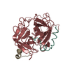
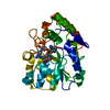
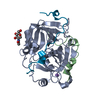
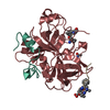
| Title | STRUCTURAL BASIS FOR SELECTIVITY OF A SMALL MOLECULE, S1-BINDING, SUB-MICROMOLAR INHIBITOR OF UROKINASE TYPE PLASMINOGEN ACTIVATOR | |||||||||
 Components Components |
| |||||||||
 Keywords Keywords | BLOOD CLOTTING/HYDROLASE INHIBITOR / selective / S1 site inhibitor / structure-based drug design / urokinase / trypsin / thrombin / BLOOD CLOTTING-HYDROLASE INHIBITOR COMPLEX | |||||||||
| Function / homology |  Function and homology information Function and homology information: / thrombospondin receptor activity / thrombin / thrombin-activated receptor signaling pathway / Defective factor XII causes hereditary angioedema / negative regulation of astrocyte differentiation / regulation of blood coagulation / neutrophil-mediated killing of gram-negative bacterium / positive regulation of phospholipase C-activating G protein-coupled receptor signaling pathway / Defective F8 cleavage by thrombin ...: / thrombospondin receptor activity / thrombin / thrombin-activated receptor signaling pathway / Defective factor XII causes hereditary angioedema / negative regulation of astrocyte differentiation / regulation of blood coagulation / neutrophil-mediated killing of gram-negative bacterium / positive regulation of phospholipase C-activating G protein-coupled receptor signaling pathway / Defective F8 cleavage by thrombin / ligand-gated ion channel signaling pathway / Platelet Aggregation (Plug Formation) / positive regulation of collagen biosynthetic process / negative regulation of platelet activation / negative regulation of blood coagulation / negative regulation of fibrinolysis / blood coagulation, fibrin clot formation / positive regulation of blood coagulation / Transport of gamma-carboxylated protein precursors from the endoplasmic reticulum to the Golgi apparatus / : / Gamma-carboxylation of protein precursors / Removal of aminoterminal propeptides from gamma-carboxylated proteins / regulation of cytosolic calcium ion concentration / fibrinolysis / : / negative regulation of proteolysis / negative regulation of cytokine production involved in inflammatory response / Regulation of Complement cascade / positive regulation of release of sequestered calcium ion into cytosol / acute-phase response / Cell surface interactions at the vascular wall / Peptide ligand-binding receptors / growth factor activity / positive regulation of receptor signaling pathway via JAK-STAT / serine-type endopeptidase inhibitor activity / lipopolysaccharide binding / platelet activation / positive regulation of protein localization to nucleus / response to wounding / Golgi lumen / Regulation of Insulin-like Growth Factor (IGF) transport and uptake by Insulin-like Growth Factor Binding Proteins (IGFBPs) / positive regulation of reactive oxygen species metabolic process / blood coagulation / positive regulation of insulin secretion / regulation of cell shape / antimicrobial humoral immune response mediated by antimicrobial peptide / heparin binding / toxin activity / Thrombin signalling through proteinase activated receptors (PARs) / positive regulation of cell growth / blood microparticle / G alpha (q) signalling events / positive regulation of phosphatidylinositol 3-kinase/protein kinase B signal transduction / cell surface receptor signaling pathway / endoplasmic reticulum lumen / receptor ligand activity / serine-type endopeptidase activity / signaling receptor binding / calcium ion binding / positive regulation of cell population proliferation / proteolysis / : / extracellular exosome / extracellular region / plasma membrane Similarity search - Function | |||||||||
| Biological species |  Homo sapiens (human) Homo sapiens (human) Hirudo medicinalis (medicinal leech) Hirudo medicinalis (medicinal leech) | |||||||||
| Method |  X-RAY DIFFRACTION / DIFFERENCE FOURIER PLUS REFINEMENT / Resolution: 1.47 Å X-RAY DIFFRACTION / DIFFERENCE FOURIER PLUS REFINEMENT / Resolution: 1.47 Å | |||||||||
 Authors Authors | Katz, B.A. / Mackman, R. / Luong, C. / Radika, K. / Martelli, A. / Sprengeler, P.A. / Wang, J. / Chan, H. / Wong, L. | |||||||||
 Citation Citation | Journal: Chem.Biol. / Year: 2000 Title: Structural basis for selectivity of a small molecule, S1-binding, submicromolar inhibitor of urokinase-type plasminogen activator. Authors: Katz, B.A. / Mackman, R. / Luong, C. / Radika, K. / Martelli, A. / Sprengeler, P.A. / Wang, J. / Chan, H. / Wong, L. | |||||||||
| History |
|
- Structure visualization
| Structure viewer | Molecule: MolmilJmol/JSmol |
|---|
- Downloads & links
-Download
| PDBx/mmCIF format | 1c5l.cif.gz | 151.3 KB | Display | PDBx/mmCIF format |
|---|---|---|---|---|
| PDB format | pdb1c5l.ent.gz | 120.7 KB | Display | PDB format |
| PDBx/mmJSON format | 1c5l.json.gz | Tree view | PDBx/mmJSON format | |
| Others |  Other downloads Other downloads |
-Validation report
| Arichive directory | https://data.pdbj.org/pub/pdb/validation_reports/c5/1c5lftp://data.pdbj.org/pub/pdb/validation_reports/c5/1c5l | HTTPS FTP |
|---|
-Related structure data
| Related structure data |  1c5mC  1c5nC  1c5oC  1c5pC  1c5qC  1c5rC  1c5sC  1c5tC  1c5uC  1c5vC  1c5wC  1c5xC  1c5yC  1c5zC C: citing same article ( |
|---|---|
| Similar structure data |


-Links
 PDBj
PDBj


- Assembly
| Deposited unit | 
| ||||||||
|---|---|---|---|---|---|---|---|---|---|
| 1 |
| ||||||||
| Unit cell |
|
-Components
-Protein/peptide , 2 types, 2 molecules LI
| #1: Protein/peptide | Mass: 4096.534 Da / Num. of mol.: 1 / Fragment: LIGHT CHAIN / Source method: isolated from a natural source / Source: (natural) Homo sapiens (human) / References: UniProt: P00734, thrombin |
|---|---|
| #3: Protein/peptide | Mass: 1363.399 Da / Num. of mol.: 1 / Source method: obtained synthetically / Source: (synth.) Hirudo medicinalis (medicinal leech) / References: UniProt: P28504, UniProt: P01050*PLUS |
-Protein , 1 types, 1 molecules H
| #2: Protein | Mass: 29780.219 Da / Num. of mol.: 1 / Fragment: HEAVY CHAIN / Source method: isolated from a natural source / Source: (natural) Homo sapiens (human) / References: UniProt: P00734, thrombin |
|---|
-Non-polymers , 3 types, 295 molecules 


| #4: Chemical | ChemComp-NA /  Mass: 22.990 Da / Num. of mol.: 1 / Source method: obtained synthetically / Formula: Na Mass: 22.990 Da / Num. of mol.: 1 / Source method: obtained synthetically / Formula: Na |
|---|---|
| #5: Chemical | ChemComp-CA /  Mass: 40.078 Da / Num. of mol.: 1 / Source method: obtained synthetically / Formula: Ca Mass: 40.078 Da / Num. of mol.: 1 / Source method: obtained synthetically / Formula: Ca |
| #6: Water | ChemComp-HOH / Mass: 18.015 Da / Num. of mol.: 293 / Source method: isolated from a natural source / Formula: H2O |
-Details
| Has protein modification | Y |
|---|
-Experimental details
-Experiment
| Experiment | Method: X-RAY DIFFRACTION / Number of used crystals: 2 |
|---|
- Sample preparation
| Crystal | Density Matthews: 2.74 Å3/Da / Density % sol: 36.7 % | |||||||||||||||||||||||||||||||||||
|---|---|---|---|---|---|---|---|---|---|---|---|---|---|---|---|---|---|---|---|---|---|---|---|---|---|---|---|---|---|---|---|---|---|---|---|---|
| Crystal grow | Method: vapor diffusion / pH: 8.2 Details: Thrombin was purchased from Haematologic Technologies, Inc. and acetyl-hirudin from Bachem. Thrombin was prepared as described (Skrzpczak-Jankun et al., 1991). Thrombin (1.0 mg/ml in 50 mM ...Details: Thrombin was purchased from Haematologic Technologies, Inc. and acetyl-hirudin from Bachem. Thrombin was prepared as described (Skrzpczak-Jankun et al., 1991). Thrombin (1.0 mg/ml in 50 mM HEPES, 50 % glycerol, pH 7.0) was incubated with 1.0 mM acetyl-hirudin for 1 hr at 4 deg C. Glycerol was removed and the complex concentrated with a centricon 10 (Amicon) to about 10 mg/ml as determined by the Biorad protein assay kit using bovine serum albumin. Crystals of thrombin-acetyl-hirudin were grown in hanging drops by vapor diffusion after streak seeding. The drops were made from 5 microliters of complex and 5 microliters of reservoir solution (0.10 M Tris, 0.50 M NaCl, 22 % (by volume) PEG 4K, pH 8.20)., VAPOR DIFFUSION | |||||||||||||||||||||||||||||||||||
| Crystal grow | *PLUS pH: 7.5 / Method: vapor diffusion, hanging drop / Details: used seeding | |||||||||||||||||||||||||||||||||||
| Components of the solutions | *PLUS
|
-Data collection
| Diffraction | Mean temperature: 298 K |
|---|---|
| Diffraction source | Source: ROTATING ANODE / Wavelength: 1.5418 |
| Detector | Type: RIGAKU RAXIS IV++ / Detector: IMAGE PLATE / Date: Apr 8, 1999 / Details: MSC MIRRORS |
| Radiation | Protocol: SINGLE WAVELENGTH / Monochromatic (M) / Laue (L): M / Scattering type: x-ray |
| Radiation wavelength | Wavelength: 1.5418 Å / Relative weight: 1 |
| Reflection | Resolution: 1.31→44.16 Å / Num. all: 51674 / % possible obs: 69 % / Observed criterion σ(I): 0.8 / Redundancy: 2.2 % / Rmerge(I) obs: 0.071 / Net I/σ(I): 9.8 |
| Reflection shell | Resolution: 1.47→1.54 Å / Rmerge(I) obs: 0.371 / Mean I/σ(I) obs: 1.3 / % possible all: 35.8 |
- Processing
| Software |
| |||||||||||||||||||||||||||
|---|---|---|---|---|---|---|---|---|---|---|---|---|---|---|---|---|---|---|---|---|---|---|---|---|---|---|---|---|
| Refinement | Method to determine structure: DIFFERENCE FOURIER PLUS REFINEMENT Resolution: 1.47→7.5 Å / Cross valid method: X-PLOR / σ(F): 1.8 Details: Met_H84, Met_H106, Met_H210, Glu_H97A, Glu_H217, Lys_H224, Ser_H27, and Glu_H192 were simultaneously refined in two conformations. No density was observed for Trp148, Thr149, Ala149A, ...Details: Met_H84, Met_H106, Met_H210, Glu_H97A, Glu_H217, Lys_H224, Ser_H27, and Glu_H192 were simultaneously refined in two conformations. No density was observed for Trp148, Thr149, Ala149A, Asn149B, Val149C, Gly149D, and Lys149E in the autolysis loop, and these residues are not included in the model. Disordered waters include: HOH395 which is in a special position; HOH396 is close to a symmetry related equivalent of itself; HOH422 which is close to HOH432; HOH434 is close to a symmetry related equivalent of itself; HOH447 which is close to HOH494; (THE ABOVE WATERS HAVE VERY STRONG DENSITY AND THEIR REFINED OCCUPANCIES ARE SIGNIFICANTLY GREATER THAN UNITY. THEY MAY REFLECT A DIFFERENT STRUCTURAL FEATURE THAN DISORDERED WATERS). HOH495 which is close to HOH499; HOH504 which is close to HOH506; HOH578 which is close to a symmetry-related equivalent of HOH579; HOH580 WHICH IS CLOSE TO HOH578 AND TO A SYMMETRY-RELATED EQUIVALENT OF HOH579; HOH652 WHICH IS CLOSE TO HOH759; HOH687 WHICH IS CLOSE TO HOH688; HOH717 WHICH IS CLOSE TO THE SIDE CHAIN OF LYS_H145; HOH733 WHICH IS CLOSE TO THE SIDE CHAIN OF SER_L1E; HOH786 IS CLOSE TO A SYMMETRY RELATED EQUIVALENT OF ITSELF. HIS_H57 IS MONOPROTONATED ON THE DELTA NITROGEN. HIS_H91 AND HIS_ H119 ARE MONOPROTONATED ON THE EPSILON NITROGEN.
| |||||||||||||||||||||||||||
| Refinement step | Cycle: LAST / Resolution: 1.47→7.5 Å
| |||||||||||||||||||||||||||
| Refine LS restraints |
| |||||||||||||||||||||||||||
| LS refinement shell | Resolution: 1.47→1.54 Å / Total num. of bins used: 8
| |||||||||||||||||||||||||||
| Xplor file |
| |||||||||||||||||||||||||||
| Software | *PLUS Name: X-PLOR / Version: 3.1 / Classification: refinement | |||||||||||||||||||||||||||
| Refinement | *PLUS Lowest resolution: 7.5 Å / σ(F): 1.8 / % reflection Rfree: 10 % / Rfactor obs: 0.217 | |||||||||||||||||||||||||||
| Solvent computation | *PLUS | |||||||||||||||||||||||||||
| Displacement parameters | *PLUS | |||||||||||||||||||||||||||
| Refine LS restraints | *PLUS
| |||||||||||||||||||||||||||
| LS refinement shell | *PLUS Rfactor Rfree: 0.532 / Rfactor Rwork: 0.527 |
