 Movie
Movie Controller

[English] 日本語
 Yorodumi
- PDB-1o3p: Elaborate Manifold of Short Hydrogen Bond Arrays Mediating Bindin... -
+ Open data

- Basic information
| Entry | Database: PDB / ID: 1o3p | ||||||
|---|---|---|---|---|---|---|---|
| Title | Elaborate Manifold of Short Hydrogen Bond Arrays Mediating Binding of Active Site-Directed Serine Protease Inhibitors | ||||||
 Components Components | (Urokinase-type plasminogen activator) x 2 | ||||||
 Keywords Keywords | BLOOD CLOTTING / hydrolase / serine protease / short hydrogen bond / inhibition mechanism / shift of pKa / trypsin / thrombin / urokinase / factor Xa | ||||||
| Function / homology |  Function and homology information Function and homology informationu-plasminogen activator / regulation of smooth muscle cell-matrix adhesion / urokinase plasminogen activator signaling pathway / regulation of plasminogen activation / regulation of integrin-mediated signaling pathway / protein complex involved in cell-matrix adhesion / regulation of fibrinolysis / regulation of wound healing / negative regulation of plasminogen activation / serine-type endopeptidase complex ...u-plasminogen activator / regulation of smooth muscle cell-matrix adhesion / urokinase plasminogen activator signaling pathway / regulation of plasminogen activation / regulation of integrin-mediated signaling pathway / protein complex involved in cell-matrix adhesion / regulation of fibrinolysis / regulation of wound healing / negative regulation of plasminogen activation / serine-type endopeptidase complex / regulation of smooth muscle cell migration / Dissolution of Fibrin Clot / regulation of cell adhesion mediated by integrin / smooth muscle cell migration / plasminogen activation / tertiary granule membrane / negative regulation of fibrinolysis / regulation of cell adhesion / serine protease inhibitor complex / specific granule membrane / fibrinolysis / positive regulation of epidermal growth factor receptor signaling pathway / chemotaxis / blood coagulation / regulation of cell population proliferation / response to hypoxia / positive regulation of cell migration / receptor ligand activity / serine-type endopeptidase activity / external side of plasma membrane / focal adhesion / Neutrophil degranulation / cell surface / signal transduction / proteolysis / : / extracellular exosome / extracellular region / plasma membrane Similarity search - Function | ||||||
| Biological species |  Homo sapiens (human) Homo sapiens (human) | ||||||
| Method |  X-RAY DIFFRACTION / FOURIER SYNTHESIS / Resolution: 1.81 Å X-RAY DIFFRACTION / FOURIER SYNTHESIS / Resolution: 1.81 Å | ||||||
 Authors Authors | Katz, B.A. / Elrod, K. / Verner, E. / Mackman, R.L. / Luong, C. / Shrader, W.D. / Sendzik, M. / Spencer, J.R. / Sprengeler, P.A. / Kolesnikov, A. ...Katz, B.A. / Elrod, K. / Verner, E. / Mackman, R.L. / Luong, C. / Shrader, W.D. / Sendzik, M. / Spencer, J.R. / Sprengeler, P.A. / Kolesnikov, A. / Tai, V.W. / Hui, H.C. / Breitenbucher, J.G. / Allen, D. / Janc, J.W. | ||||||
 Citation Citation | Journal: J.Mol.Biol. / Year: 2003 Title: Elaborate manifold of short hydrogen bond arrays mediating binding of active site-directed serine protease inhibitors. Authors: Katz, B.A. / Elrod, K. / Verner, E. / Mackman, R.L. / Luong, C. / Shrader, W.D. / Sendzik, M. / Spencer, J.R. / Sprengeler, P.A. / Kolesnikov, A. / Tai, V.W. / Hui, H.C. / Breitenbucher, J.G. ...Authors: Katz, B.A. / Elrod, K. / Verner, E. / Mackman, R.L. / Luong, C. / Shrader, W.D. / Sendzik, M. / Spencer, J.R. / Sprengeler, P.A. / Kolesnikov, A. / Tai, V.W. / Hui, H.C. / Breitenbucher, J.G. / Allen, D. / Janc, J.W. | ||||||
| History |
|
- Structure visualization
| Structure viewer | Molecule: MolmilJmol/JSmol |
|---|
- Downloads & links
-Download
| PDBx/mmCIF format | 1o3p.cif.gz | 136.6 KB | Display | PDBx/mmCIF format |
|---|---|---|---|---|
| PDB format | pdb1o3p.ent.gz | 108.5 KB | Display | PDB format |
| PDBx/mmJSON format | 1o3p.json.gz | Tree view | PDBx/mmJSON format | |
| Others |  Other downloads Other downloads |
-Validation report
| Arichive directory | https://data.pdbj.org/pub/pdb/validation_reports/o3/1o3pftp://data.pdbj.org/pub/pdb/validation_reports/o3/1o3p | HTTPS FTP |
|---|
-Related structure data
| Related structure data | 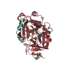 1o2gC 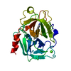 1o2hC 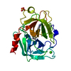 1o2iC 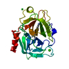 1o2jC  1o2kC  1o2lC  1o2mC  1o2nC  1o2oC  1o2pC  1o2qC  1o2rC  1o2sC  1o2tC  1o2uC  1o2vC  1o2wC  1o2xC  1o2yC  1o2zC  1o30C  1o31C  1o32C  1o33C  1o34C  1o35C  1o36C  1o37C  1o38C  1o39C  1o3aC  1o3bC  1o3cC  1o3dC  1o3eC  1o3fC  1o3gC  1o3hC  1o3iC  1o3jC  1o3kC  1o3lC  1o3mC  1o3nC  1o3oC C: citing same article ( |
|---|---|
| Similar structure data |


-Links
 PDBj
PDBj


- Assembly
| Deposited unit | 
| ||||||||||||
|---|---|---|---|---|---|---|---|---|---|---|---|---|---|
| 1 |
| ||||||||||||
| Unit cell |
| ||||||||||||
| Components on special symmetry positions |
|
-Components
| #1: Protein/peptide | Mass: 2708.183 Da / Num. of mol.: 1 / Fragment: SHORT CHAIN Source method: isolated from a genetically manipulated source Source: (gene. exp.) Homo sapiens (human) / Gene: PLAU / Plasmid: PPIC9LMWUPA / Production host:  Pichia pastoris (fungus) / References: UniProt: P00749, u-plasminogen activator Pichia pastoris (fungus) / References: UniProt: P00749, u-plasminogen activator | ||||||
|---|---|---|---|---|---|---|---|
| #2: Protein | Mass: 28435.428 Da / Num. of mol.: 1 / Fragment: CATALYTIC DOMAIN / Mutation: N145A Source method: isolated from a genetically manipulated source Source: (gene. exp.) Homo sapiens (human) / Gene: PLAU / Plasmid: PPIC9LMWUPA / Production host: Pichia pastoris (fungus) / References: UniProt: P00749, u-plasminogen activator | ||||||
| #3: Chemical |   Mass: 192.124 Da / Num. of mol.: 2 / Source method: obtained synthetically / Formula: C6H8O7 Mass: 192.124 Da / Num. of mol.: 2 / Source method: obtained synthetically / Formula: C6H8O7#4: Chemical | ChemComp-655 / |   Mass: 336.388 Da / Num. of mol.: 1 / Source method: obtained synthetically / Formula: C19H20N4O2 Mass: 336.388 Da / Num. of mol.: 1 / Source method: obtained synthetically / Formula: C19H20N4O2#5: Water | ChemComp-HOH / |  Mass: 18.015 Da / Num. of mol.: 341 / Source method: isolated from a natural source / Formula: H2O Mass: 18.015 Da / Num. of mol.: 341 / Source method: isolated from a natural source / Formula: H2OHas protein modification | Y | |
-Experimental details
-Experiment
| Experiment | Method: X-RAY DIFFRACTION / Number of used crystals: 1 |
|---|
- Sample preparation
| Crystal | Density Matthews: 2 Å3/Da / Density % sol: 38.5 % |
|---|---|
| Crystal grow | Temperature: 298 K / Method: vapor diffusion / pH: 6.5 Details: 2-propanol, PEG 4000, pH 6.5, vapor diffusion at 298 K, pH 6.50 |
-Data collection
| Diffraction | Mean temperature: 298 K |
|---|---|
| Diffraction source | Source: ROTATING ANODE / Type: RIGAKU RU200 / Wavelength: 1.5418 |
| Detector | Type: RIGAKU RAXIS IV / Detector: IMAGE PLATE / Date: Apr 11, 2000 |
| Radiation | Protocol: SINGLE WAVELENGTH / Monochromatic (M) / Laue (L): M / Scattering type: x-ray |
| Radiation wavelength | Wavelength: 1.5418 Å / Relative weight: 1 |
| Reflection | Resolution: 1.81→41.66 Å / Num. all: 22960 / Num. obs: 15430 / % possible obs: 67.2 % / Observed criterion σ(I): 0.8 / Redundancy: 2 % / Rmerge(I) obs: 0.079 / Net I/σ(I): 6.1 |
| Reflection shell | Resolution: 1.81→1.89 Å / % possible obs: 36.6 % / Rmerge(I) obs: 0.215 / Num. unique all: 2749 |
- Processing
| Software |
| ||||||||||||||||||||
|---|---|---|---|---|---|---|---|---|---|---|---|---|---|---|---|---|---|---|---|---|---|
| Refinement | Method to determine structure: FOURIER SYNTHESIS / Resolution: 1.81→7 Å / Cross valid method: THROUGHOUT / σ(F): 1.6 / Stereochemistry target values: X-PLOR force field Details: Only Leu_A9 to Lys_A16 are included for the A-chain. Residues prior and after these residues are not visible (disordered). Residues after Lys_B243 are not visible (disordered). Residues ...Details: Only Leu_A9 to Lys_A16 are included for the A-chain. Residues prior and after these residues are not visible (disordered). Residues after Lys_B243 are not visible (disordered). Residues simultaneously refined in two or more conformations are: Lys_A10, Met_B47, Glu_B84, Glu_B86, Leu_B123, Thr_B139, Gln_B192, Arg_B217, Leu_B235. HIS_H57 IS doubly protonated. HIS_H91 and His_H119 are MONOPROTONATED ON the epsilon nitrogen Disordered waters are: HOH382 which is close to conformation 1 of Arg_B217; HOH582 which is close to a symmetry-related equivalent of itself; HOH721 which is close to conformation 1 of Lys_A10; HOH1010 which is close to a symmetry-related equivalent of itself; Some of the waters may correspond to the disordered or mobile termini of the light chain. No energy terms between citrate 1 and 2 are included because they are hydrogen-bonded to one another via an unusually short hydrogen bond between carboxylate / hydroxyl groups. No energy terms are included among HOH_849, and OgSer195, and O6' of the inhibitor. These atoms form a very short multi-centered hydrogen-bonding network.
| ||||||||||||||||||||
| Refinement step | Cycle: LAST / Resolution: 1.81→7 Å
| ||||||||||||||||||||
| Refine LS restraints |
|
