 Movie
Movie Controller

[English] 日本語
 Yorodumi
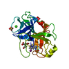
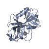
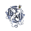
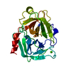
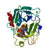
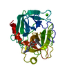
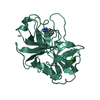
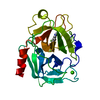
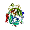
- PDB-1o2u: Elaborate Manifold of Short Hydrogen Bond Arrays Mediating Bindin... -
+ Open data

- Basic information
| Entry | Database: PDB / ID: 1o2u | ||||||
|---|---|---|---|---|---|---|---|
| Title | Elaborate Manifold of Short Hydrogen Bond Arrays Mediating Binding of Active Site-Directed Serine Protease Inhibitors | ||||||
 Components Components | BETA-TRYPSIN | ||||||
 Keywords Keywords | HYDROLASE / serine protease / short hydrogen bond / inhibition mechanism / shift of pKa / trypsin / thrombin / urokinase / factor Xa | ||||||
| Function / homology |  Function and homology information Function and homology informationtrypsin / serpin family protein binding / serine protease inhibitor complex / digestion / endopeptidase activity / serine-type endopeptidase activity / proteolysis / : / metal ion binding Similarity search - Function | ||||||
| Biological species |  | ||||||
| Method |  X-RAY DIFFRACTION / FOURIER SYNTHESIS / Resolution: 1.41 Å X-RAY DIFFRACTION / FOURIER SYNTHESIS / Resolution: 1.41 Å | ||||||
 Authors Authors | Katz, B.A. / Elrod, K. / Verner, E. / Mackman, R.L. / Luong, C. / Shrader, W.D. / Sendzik, M. / Spencer, J.R. / Sprengeler, P.A. / Kolesnikov, A. ...Katz, B.A. / Elrod, K. / Verner, E. / Mackman, R.L. / Luong, C. / Shrader, W.D. / Sendzik, M. / Spencer, J.R. / Sprengeler, P.A. / Kolesnikov, A. / Tai, V.W. / Hui, H.C. / Breitenbucher, J.G. / Allen, D. / Janc, J.W. | ||||||
 Citation Citation | Journal: J.Mol.Biol. / Year: 2003 Title: Elaborate manifold of short hydrogen bond arrays mediating binding of active site-directed serine protease inhibitors. Authors: Katz, B.A. / Elrod, K. / Verner, E. / Mackman, R.L. / Luong, C. / Shrader, W.D. / Sendzik, M. / Spencer, J.R. / Sprengeler, P.A. / Kolesnikov, A. / Tai, V.W. / Hui, H.C. / Breitenbucher, J.G. ...Authors: Katz, B.A. / Elrod, K. / Verner, E. / Mackman, R.L. / Luong, C. / Shrader, W.D. / Sendzik, M. / Spencer, J.R. / Sprengeler, P.A. / Kolesnikov, A. / Tai, V.W. / Hui, H.C. / Breitenbucher, J.G. / Allen, D. / Janc, J.W. | ||||||
| History |
|
- Structure visualization
| Structure viewer | Molecule: MolmilJmol/JSmol |
|---|
- Downloads & links
-Download
| PDBx/mmCIF format | 1o2u.cif.gz | 103.4 KB | Display | PDBx/mmCIF format |
|---|---|---|---|---|
| PDB format | pdb1o2u.ent.gz | 81.3 KB | Display | PDB format |
| PDBx/mmJSON format | 1o2u.json.gz | Tree view | PDBx/mmJSON format | |
| Others |  Other downloads Other downloads |
-Validation report
| Arichive directory | https://data.pdbj.org/pub/pdb/validation_reports/o2/1o2uftp://data.pdbj.org/pub/pdb/validation_reports/o2/1o2u | HTTPS FTP |
|---|
-Related structure data
| Related structure data | 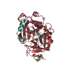 1o2gC 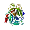 1o2hC 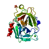 1o2iC 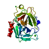 1o2jC 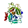 1o2kC 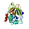 1o2lC 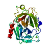 1o2mC 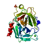 1o2nC  1o2oC 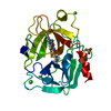 1o2pC 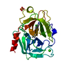 1o2qC  1o2rC  1o2sC  1o2tC 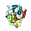 1o2vC 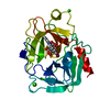 1o2wC 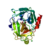 1o2xC 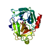 1o2yC 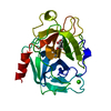 1o2zC 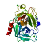 1o30C 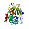 1o31C 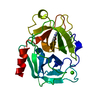 1o32C  1o33C 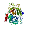 1o34C  1o35C  1o36C 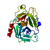 1o37C 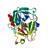 1o38C 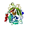 1o39C 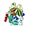 1o3aC 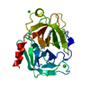 1o3bC  1o3cC 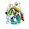 1o3dC  1o3eC  1o3fC  1o3gC 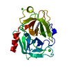 1o3hC  1o3iC 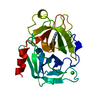 1o3jC 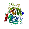 1o3kC 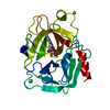 1o3lC  1o3mC 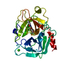 1o3nC  1o3oC  1o3pC C: citing same article ( |
|---|---|
| Similar structure data |


-Links
 PDBj
PDBj


- Assembly
| Deposited unit | 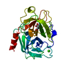
| ||||||||||
|---|---|---|---|---|---|---|---|---|---|---|---|
| 1 |
| ||||||||||
| Unit cell |
|
-Components
| #1: Protein | Mass: 23324.287 Da / Num. of mol.: 1 / Source method: isolated from a natural source / Source: (natural) |
|---|---|
| #2: Chemical | ChemComp-CA /   Mass: 40.078 Da / Num. of mol.: 1 / Source method: obtained synthetically / Formula: Ca Mass: 40.078 Da / Num. of mol.: 1 / Source method: obtained synthetically / Formula: Ca |
| #3: Chemical | ChemComp-847 / 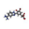  Mass: 445.224 Da / Num. of mol.: 1 / Source method: obtained synthetically / Formula: C18H13BrN4O5 Mass: 445.224 Da / Num. of mol.: 1 / Source method: obtained synthetically / Formula: C18H13BrN4O5 |
| #4: Water | ChemComp-HOH /  Mass: 18.015 Da / Num. of mol.: 136 / Source method: isolated from a natural source / Formula: H2O Mass: 18.015 Da / Num. of mol.: 136 / Source method: isolated from a natural source / Formula: H2O |
| Has protein modification | Y |
-Experimental details
-Experiment
| Experiment | Method: X-RAY DIFFRACTION / Number of used crystals: 1 |
|---|
- Sample preparation
| Crystal | Density Matthews: 2.96 Å3/Da / Density % sol: 58.51 % |
|---|---|
| Crystal grow | Temperature: 298 K / Method: vapor diffusion / pH: 8.1 Details: sodium citrate soak at target pH (10.34). vapor diffusion at 298 K, pH 8.10 |
-Data collection
| Diffraction | Mean temperature: 285 K / Ambient temp details: Crystal Cooler |
|---|---|
| Diffraction source | Source: ROTATING ANODE / Type: RIGAKU RUH3R / Wavelength: 1.5418 |
| Detector | Type: RIGAKU RAXIS IV++ / Detector: IMAGE PLATE / Date: Mar 25, 2002 |
| Radiation | Protocol: SINGLE WAVELENGTH / Monochromatic (M) / Laue (L): M / Scattering type: x-ray |
| Radiation wavelength | Wavelength: 1.5418 Å / Relative weight: 1 |
| Reflection | Resolution: 1.41→23.59 Å / Num. all: 54164 / Num. obs: 49944 / % possible obs: 92 % / Observed criterion σ(I): 0 / Redundancy: 2.6 % / Rmerge(I) obs: 0.033 / Net I/σ(I): 11.5 |
| Reflection shell | Resolution: 1.41→1.47 Å / % possible obs: 51.3 % / Rmerge(I) obs: 0.317 / Mean I/σ(I) obs: 0.9 / Num. unique all: 6659 |
- Processing
| Software |
| ||||||||||||||||||||
|---|---|---|---|---|---|---|---|---|---|---|---|---|---|---|---|---|---|---|---|---|---|
| Refinement | Method to determine structure: FOURIER SYNTHESIS / Resolution: 1.41→7 Å / Cross valid method: THROUGHOUT / σ(F): 0.5 / Stereochemistry target values: X-PLOR force field Details: Residues simultaneously refined in two or more conformations are: Val53, Leu66, Ser86, Lys87, Ser110, Ser113, Ser130, Lys159, Asp165, Ser170, Gln175, Ser195, Ser217, Lys230, Ser236, Ser244, ...Details: Residues simultaneously refined in two or more conformations are: Val53, Leu66, Ser86, Lys87, Ser110, Ser113, Ser130, Lys159, Asp165, Ser170, Gln175, Ser195, Ser217, Lys230, Ser236, Ser244, Bzm246 HOH383 makes a short H-bond with 847246 (conf 1). Other H-bonds are normal. His40 and HIS91 are MONOPROTONATED ON THE EPSILON NITROGEN.
| ||||||||||||||||||||
| Refinement step | Cycle: LAST / Resolution: 1.41→7 Å
| ||||||||||||||||||||
| Refine LS restraints |
|
