 Movie
Movie Controller


+ Open data

- Basic information
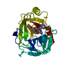
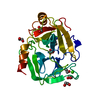
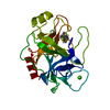
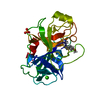
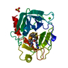
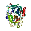
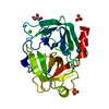
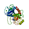
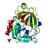
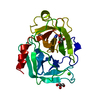
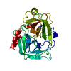
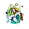
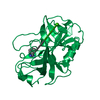
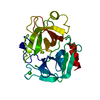
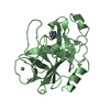
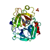
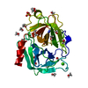
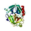
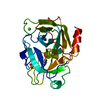
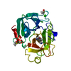
| Entry | Database: PDB / ID: 2zdk | ||||||
|---|---|---|---|---|---|---|---|
| Title | Exploring Trypsin S3 Pocket | ||||||
 Components Components | Cationic trypsin | ||||||
 Keywords Keywords | Hydrolase/Hydrolase Inhibitor / Hydrolase Inhibitors / Digestion / Metal-binding / Protease / Secreted / Serine protease / Zymogen / Hydrolase-Hydrolase Inhibitor complex | ||||||
| Function / homology |  Function and homology information Function and homology informationtrypsin / serpin family protein binding / serine protease inhibitor complex / digestion / endopeptidase activity / serine-type endopeptidase activity / proteolysis / : / metal ion binding Similarity search - Function | ||||||
| Biological species |  | ||||||
| Method |  X-RAY DIFFRACTION / MOLECULAR REPLACEMENT / Resolution: 1.67 Å X-RAY DIFFRACTION / MOLECULAR REPLACEMENT / Resolution: 1.67 Å | ||||||
 Authors Authors | Baum, B. / Brandt, T. / Heine, A. / Klebe, G. | ||||||
 Citation Citation | Journal: J.Mol.Biol. / Year: 2011 Title: Congeneric but still distinct: how closely related trypsin ligands exhibit different thermodynamic and structural properties Authors: Brandt, T. / Holzmann, N. / Muley, L. / Khayat, M. / Wegscheid-Gerlach, C. / Baum, B. / Heine, A. / Hangauer, D. / Klebe, G. | ||||||
| History |
|
- Structure visualization
| Structure viewer | Molecule: MolmilJmol/JSmol |
|---|
- Downloads & links
-Download
| PDBx/mmCIF format | 2zdk.cif.gz | 64.4 KB | Display | PDBx/mmCIF format |
|---|---|---|---|---|
| PDB format | pdb2zdk.ent.gz | 44.8 KB | Display | PDB format |
| PDBx/mmJSON format | 2zdk.json.gz | Tree view | PDBx/mmJSON format | |
| Others |  Other downloads Other downloads |
-Validation report
| Arichive directory | https://data.pdbj.org/pub/pdb/validation_reports/zd/2zdkftp://data.pdbj.org/pub/pdb/validation_reports/zd/2zdk | HTTPS FTP |
|---|
-Related structure data
| Related structure data |  2zdlC  2zdmC  2zdnC  2zfsC  2zftC  2zhdC  2zq1C  2zq2C  3ljjC  3ljoC 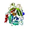 1gi1S C: citing same article ( S: Starting model for refinement |
|---|---|
| Similar structure data |
-Links
 PDBj
PDBj

- Assembly
| Deposited unit | 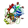
| ||||||||
|---|---|---|---|---|---|---|---|---|---|
| 1 |
| ||||||||
| Unit cell |
| ||||||||
| Components on special symmetry positions |
|
-Components
-Protein , 1 types, 1 molecules A
| #1: Protein | Mass: 23324.287 Da / Num. of mol.: 1 / Source method: isolated from a natural source / Source: (natural) |
|---|
-Non-polymers , 6 types, 240 molecules 


| #2: Chemical | ChemComp-CA /  Mass: 40.078 Da / Num. of mol.: 1 / Source method: obtained synthetically / Formula: Ca Mass: 40.078 Da / Num. of mol.: 1 / Source method: obtained synthetically / Formula: Ca | ||
|---|---|---|---|
| #3: Chemical | ChemComp-SO4 /  Mass: 96.063 Da / Num. of mol.: 1 / Source method: obtained synthetically / Formula: SO4 Mass: 96.063 Da / Num. of mol.: 1 / Source method: obtained synthetically / Formula: SO4 | ||
| #4: Chemical | ChemComp-50U / ( Type: peptide-like, Peptide-like / Class: Thrombin inhibitor / Mass: 384.515 Da / Num. of mol.: 1 / Source method: obtained synthetically / Formula: C22H32N4O2 Type: peptide-like, Peptide-like / Class: Thrombin inhibitor / Mass: 384.515 Da / Num. of mol.: 1 / Source method: obtained synthetically / Formula: C22H32N4O2References: (S)-N-(4-carbamimidoylbenzyl)-1-(3-cyclohexylpropanoyl)pyrrolidine-2-carboxamide | ||
| #5: Chemical | ChemComp-DMS /  Mass: 78.133 Da / Num. of mol.: 1 / Source method: obtained synthetically / Formula: C2H6OS / Comment: DMSO, precipitant*YM Mass: 78.133 Da / Num. of mol.: 1 / Source method: obtained synthetically / Formula: C2H6OS / Comment: DMSO, precipitant*YM | ||
| #6: Chemical | ChemComp-GOL /  Mass: 92.094 Da / Num. of mol.: 7 / Source method: obtained synthetically / Formula: C3H8O3 Mass: 92.094 Da / Num. of mol.: 7 / Source method: obtained synthetically / Formula: C3H8O3#7: Water | ChemComp-HOH / | Mass: 18.015 Da / Num. of mol.: 229 / Source method: isolated from a natural source / Formula: H2O |
-Details
| Has protein modification | Y |
|---|
-Experimental details
-Experiment
| Experiment | Method: X-RAY DIFFRACTION / Number of used crystals: 1 |
|---|
- Sample preparation
| Crystal | Density Matthews: 2 Å3/Da / Density % sol: 38.61 % |
|---|---|
| Crystal grow | Temperature: 289 K / Method: vapor diffusion, sitting drop / pH: 7 Details: 0.1M Imidazole buffer, 0.1M ammonium sulfate, 25% PEG 8000, pH 7.0, VAPOR DIFFUSION, SITTING DROP, temperature 289K |
-Data collection
| Diffraction | Mean temperature: 113 K |
|---|---|
| Diffraction source | Source: ROTATING ANODE / Type: RIGAKU RUH3R / Wavelength: 1.5418 Å |
| Detector | Type: RIGAKU RAXIS IV / Detector: IMAGE PLATE / Date: Oct 1, 2007 |
| Radiation | Protocol: SINGLE WAVELENGTH / Monochromatic (M) / Laue (L): M / Scattering type: x-ray |
| Radiation wavelength | Wavelength: 1.5418 Å / Relative weight: 1 |
| Reflection | Resolution: 1.67→30 Å / Num. all: 22193 / Num. obs: 22193 / % possible obs: 98.7 % / Redundancy: 6.2 % / Biso Wilson estimate: 15.9 Å2 / Rsym value: 0.059 / Net I/σ(I): 31.7 |
| Reflection shell | Resolution: 1.67→1.7 Å / Redundancy: 3.5 % / Mean I/σ(I) obs: 2.9 / Num. unique all: 944 / Rsym value: 0.356 / % possible all: 83.8 |
- Processing
| Software |
| |||||||||||||||||||||||||||||||||
|---|---|---|---|---|---|---|---|---|---|---|---|---|---|---|---|---|---|---|---|---|---|---|---|---|---|---|---|---|---|---|---|---|---|---|
| Refinement | Method to determine structure: MOLECULAR REPLACEMENT Starting model: PDB entry 1GI1 Resolution: 1.67→10 Å / Num. parameters: 7832 / Num. restraintsaints: 7161 / Cross valid method: FREE R / σ(F): 0 / σ(I): 0 / Stereochemistry target values: ENGH AND HUBER
| |||||||||||||||||||||||||||||||||
| Refine analyze | Num. disordered residues: 8 / Occupancy sum hydrogen: 1566 / Occupancy sum non hydrogen: 1921.5 | |||||||||||||||||||||||||||||||||
| Refinement step | Cycle: LAST / Resolution: 1.67→10 Å
| |||||||||||||||||||||||||||||||||
| Refine LS restraints |
|
