 Movie
Movie Controller

[English] 日本語
 Yorodumi
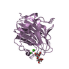
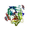
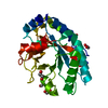
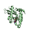
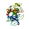
- PDB-1f2s: CRYSTAL STRUCTURE OF THE COMPLEX FORMED BETWEEN BOVINE BETA-TRYPS... -
+ Open data

- Basic information
| Entry | Database: PDB / ID: 1f2s | |||||||||
|---|---|---|---|---|---|---|---|---|---|---|
| Title | CRYSTAL STRUCTURE OF THE COMPLEX FORMED BETWEEN BOVINE BETA-TRYPSIN AND MCTI-A, A TRYPSIN INHIBITOR OF SQUASH FAMILY AT 1.8 A RESOLUTION | |||||||||
 Components Components |
| |||||||||
 Keywords Keywords | HYDROLASE/HYDROLASE INHIBITOR / PROTEINASE-INHIBITOR COMPLEX / TRYPSIN / HYDROLASE-HYDROLASE INHIBITOR COMPLEX | |||||||||
| Function / homology |  Function and homology information Function and homology informationtrypsin / serpin family protein binding / serine protease inhibitor complex / digestion / serine-type endopeptidase inhibitor activity / endopeptidase activity / serine-type endopeptidase activity / proteolysis / : / extracellular region / metal ion binding Similarity search - Function | |||||||||
| Biological species |   Momordica charantia (bitter melon) Momordica charantia (bitter melon) | |||||||||
| Method |  X-RAY DIFFRACTION / MOLECULAR REPLACEMENT / Resolution: 1.79 Å X-RAY DIFFRACTION / MOLECULAR REPLACEMENT / Resolution: 1.79 Å | |||||||||
 Authors Authors | Zhu, Y. / Huang, Q. / Qian, M. / Jia, Y. / Tang, Y. | |||||||||
 Citation Citation | Journal: J.Protein Chem. / Year: 1999 Title: Crystal structure of the complex formed between bovine beta-trypsin and MCTI-A, a trypsin inhibitor of squash family, at 1.8-A resolution. Authors: Zhu, Y. / Huang, Q. / Qian, M. / Jia, Y. / Tang, Y. #1: Journal: J.Mol.Biol. / Year: 1993Title: REFINED 1.6A RESOLUTION CRYSTAL STRUCTURE OF THE COMPLEX FORMED BETWEEN PORCINE BETA-TRYPSIN AND MCTI-A, A TRYPSIN INHIBITOR OF THE SQUASH FAMILY Authors: Huang, Q. / Liu, S. / Tang, Y. | |||||||||
| History |
|
- Structure visualization
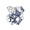
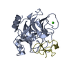
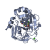
| Structure viewer | Molecule: MolmilJmol/JSmol |
|---|
- Downloads & links
-Download
| PDBx/mmCIF format | 1f2s.cif.gz | 64.6 KB | Display | PDBx/mmCIF format |
|---|---|---|---|---|
| PDB format | pdb1f2s.ent.gz | 45.8 KB | Display | PDB format |
| PDBx/mmJSON format | 1f2s.json.gz | Tree view | PDBx/mmJSON format | |
| Others |  Other downloads Other downloads |
-Validation report
| Arichive directory | https://data.pdbj.org/pub/pdb/validation_reports/f2/1f2sftp://data.pdbj.org/pub/pdb/validation_reports/f2/1f2s | HTTPS FTP |
|---|
-Related structure data
| Related structure data | 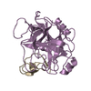 1ppeS S: Starting model for refinement |
|---|---|
| Similar structure data |


-Links
 PDBj
PDBj

- Assembly
| Deposited unit | 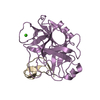
| ||||||||
|---|---|---|---|---|---|---|---|---|---|
| 1 |
| ||||||||
| Unit cell |
|
-Components
| #1: Protein | Mass: 23324.287 Da / Num. of mol.: 1 / Source method: isolated from a natural source / Details: PANCREATIC / Source: (natural) |
|---|---|
| #2: Protein/peptide | Mass: 3251.958 Da / Num. of mol.: 1 / Source method: isolated from a natural source / Details: SEED / Source: (natural) Momordica charantia (bitter melon) / References: UniProt: P10295 |
| #3: Chemical | ChemComp-CA /   Mass: 40.078 Da / Num. of mol.: 1 / Source method: obtained synthetically / Formula: Ca Mass: 40.078 Da / Num. of mol.: 1 / Source method: obtained synthetically / Formula: Ca |
| #4: Water | ChemComp-HOH /  Mass: 18.015 Da / Num. of mol.: 207 / Source method: isolated from a natural source / Formula: H2O Mass: 18.015 Da / Num. of mol.: 207 / Source method: isolated from a natural source / Formula: H2O |
| Has protein modification | Y |
-Experimental details
-Experiment
| Experiment | Method: X-RAY DIFFRACTION / Number of used crystals: 2 |
|---|
- Sample preparation
| Crystal | Density Matthews: 2.32 Å3/Da / Density % sol: 47.09 % | |||||||||||||||||||||||||||||||||||||||||||||||||
|---|---|---|---|---|---|---|---|---|---|---|---|---|---|---|---|---|---|---|---|---|---|---|---|---|---|---|---|---|---|---|---|---|---|---|---|---|---|---|---|---|---|---|---|---|---|---|---|---|---|---|
| Crystal grow | Temperature: 295 K / Method: vapor diffusion, hanging drop / pH: 5.1 Details: PEG6000, soduim phosphate buffer, pH 5.10, VAPOR DIFFUSION, HANGING DROP, temperature 295K | |||||||||||||||||||||||||||||||||||||||||||||||||
| Crystal grow | *PLUS Temperature: 22 ℃ / pH: 5 Details: drop consists of equal volume of protein and precipitant solutions | |||||||||||||||||||||||||||||||||||||||||||||||||
| Components of the solutions | *PLUS
|
-Data collection
| Diffraction | Mean temperature: 297 K |
|---|---|
| Diffraction source | Source: ROTATING ANODE / Type: RIGAKU / Wavelength: 1.54178 |
| Detector | Type: RIGAKU RAXIS IIC / Detector: IMAGE PLATE / Date: Oct 26, 1998 |
| Radiation | Protocol: SINGLE WAVELENGTH / Monochromatic (M) / Laue (L): M / Scattering type: x-ray |
| Radiation wavelength | Wavelength: 1.54178 Å / Relative weight: 1 |
| Reflection | Resolution: 1.79→75 Å / Num. all: 23032 / Num. obs: 20741 / % possible obs: 86.3 % / Observed criterion σ(F): 2 / Observed criterion σ(I): 1 / Redundancy: 5.7 % / Biso Wilson estimate: 20.9 Å2 / Rmerge(I) obs: 0.082 / Net I/σ(I): 8.7 |
| Reflection shell | Resolution: 1.79→1.86 Å / Redundancy: 4.7 % / Rmerge(I) obs: 0.213 / Num. unique all: 1607 / % possible all: 63.4 |
| Reflection shell | *PLUS % possible obs: 63.4 % |
- Processing
| Software |
| |||||||||||||||||||||||||
|---|---|---|---|---|---|---|---|---|---|---|---|---|---|---|---|---|---|---|---|---|---|---|---|---|---|---|
| Refinement | Method to determine structure: MOLECULAR REPLACEMENT Starting model: 1PPE Resolution: 1.79→7 Å / σ(F): 2 / σ(I): 1 / Stereochemistry target values: Engh & Huber
| |||||||||||||||||||||||||
| Refinement step | Cycle: LAST / Resolution: 1.79→7 Å
| |||||||||||||||||||||||||
| Refine LS restraints |
| |||||||||||||||||||||||||
| Xplor file |
| |||||||||||||||||||||||||
| Software | *PLUS Name: X-PLOR / Version: 3.1 / Classification: refinement | |||||||||||||||||||||||||
| Refine LS restraints | *PLUS
|
