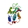
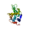
 Movie
Movie Controller


+ Open data

- Basic information
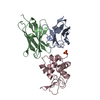
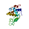
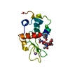
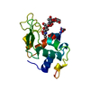
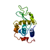
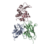
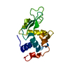
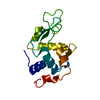
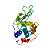
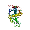
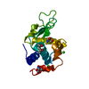
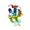
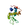
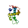
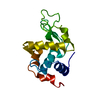
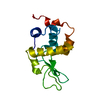
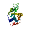
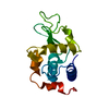
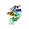
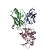
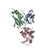
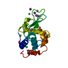
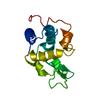
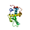
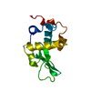
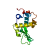
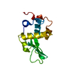
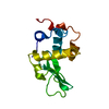
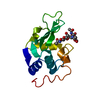
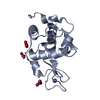
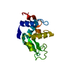
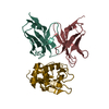
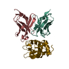
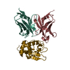
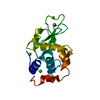
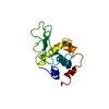
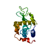
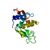
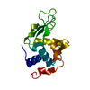
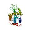
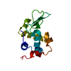
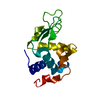
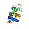
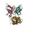
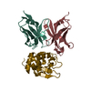
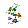
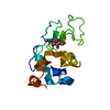
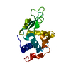
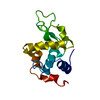
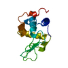
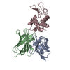
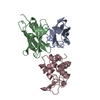
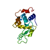
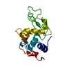
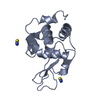
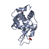
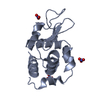
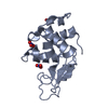
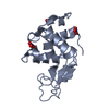
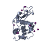
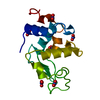
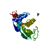
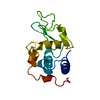
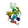
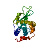
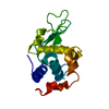
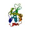
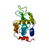
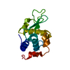
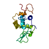
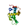
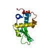
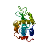
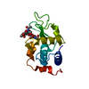
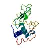
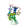
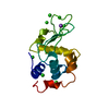
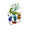
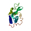
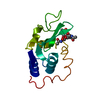
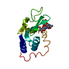
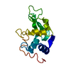
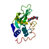
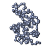
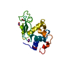
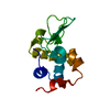
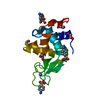
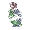
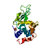
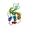
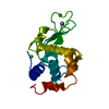
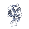
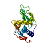
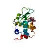
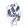
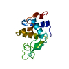
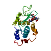
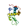
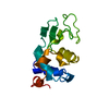
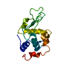
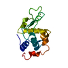
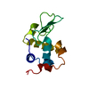
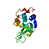
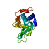
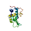
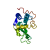
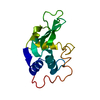
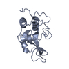
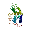
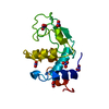
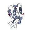
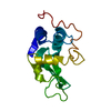
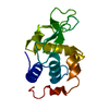
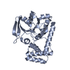
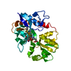
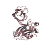
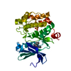
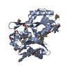
| Entry | Database: PDB / ID: 1uuz | |||||||||
|---|---|---|---|---|---|---|---|---|---|---|
| Title | IVY:A NEW FAMILY OF PROTEIN | |||||||||
 Components Components |
| |||||||||
 Keywords Keywords | HYDROLASE/INHIBITOR / LYSOZYME-INHIBITOR COMPLEX / IVY / TYPE-C LYSOZYME INHIBITOR / LYSOZYME / HYDROLASE / GLYCOSIDASE / HYDROLASE-INHIBITOR complex | |||||||||
| Function / homology |  Function and homology information Function and homology informationLactose synthesis / Antimicrobial peptides / Neutrophil degranulation / beta-N-acetylglucosaminidase activity / cell wall macromolecule catabolic process / lysozyme / lysozyme activity / killing of cells of another organism / defense response to Gram-negative bacterium / periplasmic space ...Lactose synthesis / Antimicrobial peptides / Neutrophil degranulation / beta-N-acetylglucosaminidase activity / cell wall macromolecule catabolic process / lysozyme / lysozyme activity / killing of cells of another organism / defense response to Gram-negative bacterium / periplasmic space / defense response to bacterium / defense response to Gram-positive bacterium / endoplasmic reticulum / : / identical protein binding / cytoplasm Similarity search - Function | |||||||||
| Biological species |   PSEUDOMONAS AERUGINOSA (bacteria) PSEUDOMONAS AERUGINOSA (bacteria) | |||||||||
| Method |  X-RAY DIFFRACTION / SYNCHROTRON / MOLECULAR REPLACEMENT / Resolution: 1.8 Å X-RAY DIFFRACTION / SYNCHROTRON / MOLECULAR REPLACEMENT / Resolution: 1.8 Å | |||||||||
 Authors Authors | Abergel, C. / Lembo, F. / Byrne, D. / Maza, C. / Claverie, J.M. | |||||||||
 Citation Citation | Journal: Proc.Natl.Acad.Sci.USA / Year: 2007 Title: Structure and Evolution of the Ivy Protein Family, Unexpected Lysozyme Inhibitors in Gram-Negative Bacteria. Authors: Abergel, C. / Monchois, V. / Byrne, D. / Chenivesse, S. / Lembo, F. / Lazzaroni, J.-C. / Claverie, J.M. | |||||||||
| History |
|
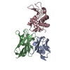
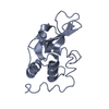
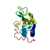
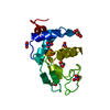
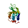
- Structure visualization
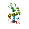
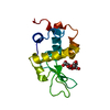
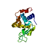
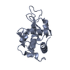
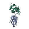
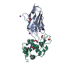
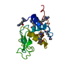
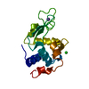
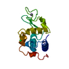
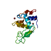
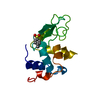
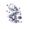
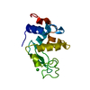
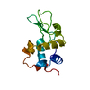
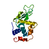
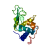
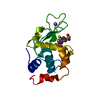
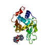
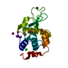
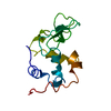
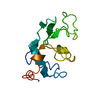
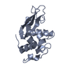
| Structure viewer | Molecule: MolmilJmol/JSmol |
|---|
- Downloads & links
-Download
| PDBx/mmCIF format | 1uuz.cif.gz | 120.6 KB | Display | PDBx/mmCIF format |
|---|---|---|---|---|
| PDB format | pdb1uuz.ent.gz | 95.5 KB | Display | PDB format |
| PDBx/mmJSON format | 1uuz.json.gz | Tree view | PDBx/mmJSON format | |
| Others |  Other downloads Other downloads |
-Validation report
| Arichive directory | https://data.pdbj.org/pub/pdb/validation_reports/uu/1uuzftp://data.pdbj.org/pub/pdb/validation_reports/uu/1uuz | HTTPS FTP |
|---|
-Related structure data
| Related structure data | 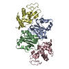 1gpqSC  1xs0C S: Starting model for refinement C: citing same article ( |
|---|---|
| Similar structure data |


-Links
 PDBj
PDBj


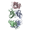
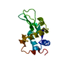
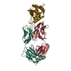
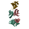
- Assembly
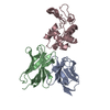
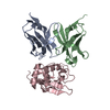
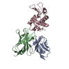
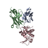
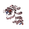
| Deposited unit | 
| ||||||||
|---|---|---|---|---|---|---|---|---|---|
| 1 | 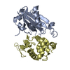
| ||||||||
| 2 | 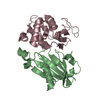
| ||||||||
| Unit cell |
|
-Components
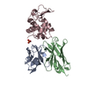
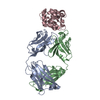
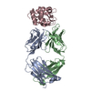
| #1: Protein | Mass: 15549.329 Da / Num. of mol.: 2 Source method: isolated from a genetically manipulated source Details: IVY COMPLEXED WITH HEWL IN CRYSTAL / Source: (gene. exp.) PSEUDOMONAS AERUGINOSA (bacteria) / Description: C-TERM HIS-TAG / Plasmid: PET26 / Production host: #2: Protein | Mass: 14331.160 Da / Num. of mol.: 2 / Source method: isolated from a natural source / Details: SIGMA / Source: (natural) #3: Water | ChemComp-HOH / |  Mass: 18.015 Da / Num. of mol.: 386 / Source method: isolated from a natural source / Formula: H2O Mass: 18.015 Da / Num. of mol.: 386 / Source method: isolated from a natural source / Formula: H2OCompound details | THE LOOP CKPHDC IS RESPONSIBL | Has protein modification | Y | |
|---|
-Experimental details
-Experiment
| Experiment | Method: X-RAY DIFFRACTION / Number of used crystals: 1 |
|---|
- Sample preparation
| Crystal | Density Matthews: 2.13 Å3/Da / Density % sol: 42.33 % | ||||||||||||||||||||||||||||||||||||
|---|---|---|---|---|---|---|---|---|---|---|---|---|---|---|---|---|---|---|---|---|---|---|---|---|---|---|---|---|---|---|---|---|---|---|---|---|---|
| Crystal grow | pH: 6.5 Details: 20% POLYETHYLENEGLYCOL 4000, IMIDAZOLE/MALATE 0.2M PH 6.0, 5% GLYCEROL | ||||||||||||||||||||||||||||||||||||
| Crystal grow | *PLUS pH: 8 / Method: vapor diffusion | ||||||||||||||||||||||||||||||||||||
| Components of the solutions | *PLUS
|
-Data collection
| Diffraction | Mean temperature: 100 K |
|---|---|
| Diffraction source | Source: SYNCHROTRON / Site: ESRF  / Beamline: ID14-2 / Wavelength: 0.933 / Beamline: ID14-2 / Wavelength: 0.933 |
| Detector | Type: ADSC CCD / Detector: CCD / Date: Sep 15, 2001 |
| Radiation | Protocol: SINGLE WAVELENGTH / Monochromatic (M) / Laue (L): M / Scattering type: x-ray |
| Radiation wavelength | Wavelength: 0.933 Å / Relative weight: 1 |
| Reflection | Resolution: 1.8→47.673 Å / Num. obs: 42990 / % possible obs: 96.5 % / Redundancy: 2.8 % / Biso Wilson estimate: 18.5 Å2 / Rmerge(I) obs: 0.057 / Net I/σ(I): 7.7 |
| Reflection shell | Resolution: 1.8→1.86 Å / Redundancy: 2.8 % / Rmerge(I) obs: 0.323 / Mean I/σ(I) obs: 2.3 / % possible all: 96.5 |
| Reflection | *PLUS Highest resolution: 1.8 Å / Lowest resolution: 25 Å / Redundancy: 2.8 % / Num. measured all: 122164 / Rmerge(I) obs: 0.057 |
| Reflection shell | *PLUS Highest resolution: 1.8 Å / % possible obs: 96.5 % / Redundancy: 2.8 % / Num. unique obs: 4166 / Num. measured obs: 11604 / Rmerge(I) obs: 0.323 / Mean I/σ(I) obs: 2.3 |
- Processing
| Software |
| ||||||||||||||||||||||||||||||||||||||||||||||||||||||||||||||||||||||||||||||||
|---|---|---|---|---|---|---|---|---|---|---|---|---|---|---|---|---|---|---|---|---|---|---|---|---|---|---|---|---|---|---|---|---|---|---|---|---|---|---|---|---|---|---|---|---|---|---|---|---|---|---|---|---|---|---|---|---|---|---|---|---|---|---|---|---|---|---|---|---|---|---|---|---|---|---|---|---|---|---|---|---|---|
| Refinement | Method to determine structure: MOLECULAR REPLACEMENT Starting model: PDB ENTRY 1GPQ Resolution: 1.8→23.94 Å / Rfactor Rfree error: 0.004 / Isotropic thermal model: RESTRAINED / Cross valid method: THROUGHOUT / σ(F): 0
| ||||||||||||||||||||||||||||||||||||||||||||||||||||||||||||||||||||||||||||||||
| Solvent computation | Solvent model: FLAT MODEL / Bsol: 37.456 Å2 / ksol: 0.325439 e/Å3 | ||||||||||||||||||||||||||||||||||||||||||||||||||||||||||||||||||||||||||||||||
| Displacement parameters | Biso mean: 27.1 Å2
| ||||||||||||||||||||||||||||||||||||||||||||||||||||||||||||||||||||||||||||||||
| Refine analyze |
| ||||||||||||||||||||||||||||||||||||||||||||||||||||||||||||||||||||||||||||||||
| Refinement step | Cycle: LAST / Resolution: 1.8→23.94 Å
| ||||||||||||||||||||||||||||||||||||||||||||||||||||||||||||||||||||||||||||||||
| Refine LS restraints |
| ||||||||||||||||||||||||||||||||||||||||||||||||||||||||||||||||||||||||||||||||
| LS refinement shell | Resolution: 1.8→1.91 Å / Rfactor Rfree error: 0.012 / Total num. of bins used: 6
| ||||||||||||||||||||||||||||||||||||||||||||||||||||||||||||||||||||||||||||||||
| Xplor file |
| ||||||||||||||||||||||||||||||||||||||||||||||||||||||||||||||||||||||||||||||||
| Refinement | *PLUS Lowest resolution: 20 Å / % reflection Rfree: 9.9 % / Rfactor Rfree: 0.25 | ||||||||||||||||||||||||||||||||||||||||||||||||||||||||||||||||||||||||||||||||
| Solvent computation | *PLUS | ||||||||||||||||||||||||||||||||||||||||||||||||||||||||||||||||||||||||||||||||
| Displacement parameters | *PLUS | ||||||||||||||||||||||||||||||||||||||||||||||||||||||||||||||||||||||||||||||||
| Refine LS restraints | *PLUS
|
