 Movie
Movie Controller

[English] 日本語
 Yorodumi
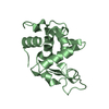
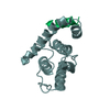
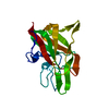
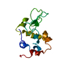
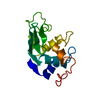
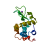
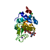
- PDB-1ja2: BINDING OF N-ACETYLGLUCOSAMINE TO CHICKEN EGG LYSOZYME: A POWDER ... -
+ Open data

- Basic information
| Entry | Database: PDB / ID: 1ja2 | ||||||
|---|---|---|---|---|---|---|---|
| Title | BINDING OF N-ACETYLGLUCOSAMINE TO CHICKEN EGG LYSOZYME: A POWDER DIFFRACTION STUDY | ||||||
 Components Components | LYSOZYME | ||||||
 Keywords Keywords | HYDROLASE / POWDER DIFFRACTION / RIETVELD REFINEMENT / LYSOZYME | ||||||
| Function / homology |  Function and homology information Function and homology informationLactose synthesis / Antimicrobial peptides / Neutrophil degranulation / beta-N-acetylglucosaminidase activity / cell wall macromolecule catabolic process / lysozyme / lysozyme activity / killing of cells of another organism / defense response to Gram-negative bacterium / defense response to bacterium ...Lactose synthesis / Antimicrobial peptides / Neutrophil degranulation / beta-N-acetylglucosaminidase activity / cell wall macromolecule catabolic process / lysozyme / lysozyme activity / killing of cells of another organism / defense response to Gram-negative bacterium / defense response to bacterium / defense response to Gram-positive bacterium / endoplasmic reticulum / : / identical protein binding / cytoplasm Similarity search - Function | ||||||
| Biological species |  | ||||||
| Method | POWDER DIFFRACTION /  SYNCHROTRON / MOLECULAR REPLACEMENT / Resolution: 2.87 Å SYNCHROTRON / MOLECULAR REPLACEMENT / Resolution: 2.87 Å | ||||||
 Authors Authors | Von Dreele, R.B. | ||||||
 Citation Citation | Journal: Acta Crystallogr.,Sect.D / Year: 2001 Title: Binding of N-acetylglucosamine to chicken egg lysozyme: a powder diffraction study. Authors: Von Dreele, R.B. #1: Journal: J.Appl.Crystallogr. / Year: 1999Title: Combined Rietveld and Stereochemical Restraint Refinement of a Protein Crystal Structure Authors: Von Dreele, R.B. #2: Journal: Acta Crystallogr.,Sect.D / Year: 2000Title: The First Protein Crystal Structure Determined from Resolution X-Ray Powder Diffraction Data: A Variant of the T3R3 Human Insulin Zinc Complex Produced by Grinding Authors: Von Dreele, R.B. / Stephens, P.W. / Blessing, R.H. / Smith, G.D. | ||||||
| History |
|
- Structure visualization
| Structure viewer | Molecule: MolmilJmol/JSmol |
|---|
- Downloads & links
-Download
| PDBx/mmCIF format | 1ja2.cif.gz | 33.1 KB | Display | PDBx/mmCIF format |
|---|---|---|---|---|
| PDB format | pdb1ja2.ent.gz | 19.7 KB | Display | PDB format |
| PDBx/mmJSON format | 1ja2.json.gz | Tree view | PDBx/mmJSON format | |
| Others |  Other downloads Other downloads |
-Validation report
| Arichive directory | https://data.pdbj.org/pub/pdb/validation_reports/ja/1ja2ftp://data.pdbj.org/pub/pdb/validation_reports/ja/1ja2 | HTTPS FTP |
|---|
-Related structure data
| Related structure data |  1ja4C  1ja6C  1ja7C  1rfpS C: citing same article ( S: Starting model for refinement |
|---|---|
| Similar structure data |


-Links
 PDBj
PDBj


- Assembly
| Deposited unit | 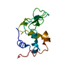
|
|---|---|
| 1 |
|
-Components
| #1: Protein | Mass: 14331.160 Da / Num. of mol.: 1 Source method: isolated from a genetically manipulated source Source: (gene. exp.)  |
|---|---|
| Has protein modification | Y |
-Experimental details
-Experiment
| Experiment | Method: POWDER DIFFRACTION / Number of used crystals: 1 |
|---|
- Sample preparation
| Crystal | Density Matthews: 2.07 Å3/Da / Density % sol: 40.68 % |
|---|---|
| Crystal grow | Temperature: 296 K / Method: precipitation / pH: 4.8 Details: rapid precipitation from 0.5M NACL IN PH 5.0 0.05M KHPHTHALATE/NAOH BUFFER, at 296K, pH 4.8 |
| Crystal grow | *PLUS Method: unknown / Details: Von Dreele, R.B., (2000) Acta Cryst., D56, 1549. |
| Components of the solutions | *PLUS Conc.: 1.0 M / Chemical formula: NaCl |
-Data collection
| Diffraction | Mean temperature: 296 K |
|---|---|
| Diffraction source | Source: SYNCHROTRON / Site: NSLS  / Beamline: X3B1 / Wavelength: 0.70003 Å / Beamline: X3B1 / Wavelength: 0.70003 Å |
| Detector | Detector: SCINTILLATOR / Date: Oct 23, 2000 / Details: 2MM X 8MM BEAM |
| Radiation | Monochromator: DOUBLE SI(111) / Protocol: SINGLE WAVELENGTH / Monochromatic (M) / Laue (L): M |
| Radiation wavelength | Wavelength: 0.70003 Å / Relative weight: 1 |
| Reflection | Resolution: 2.87→40.03 Å / Num. all: 3112 / Num. obs: 3112 / % possible obs: 100 % |
- Processing
| Software |
| ||||||||||||
|---|---|---|---|---|---|---|---|---|---|---|---|---|---|
| Refinement | Method to determine structure: MOLECULAR REPLACEMENT Starting model: PDB ENTRY 1RFP Resolution: 2.87→40.03 Å / Isotropic thermal model: Overall fixed / Stereochemistry target values: Engh & Huber Details: Used band matrix least squares, band width 300 parameters
| ||||||||||||
| Displacement parameters | Biso mean: 23.7 Å2 | ||||||||||||
| Refinement step | Cycle: LAST / Resolution: 2.87→40.03 Å
| ||||||||||||
| Refine LS restraints |
|
