 Movie
Movie Controller

[English] 日本語
 Yorodumi
- PDB-1f10: CRYSTAL STRUCTURE OF ORTHORHOMBIC LYSOZYME GROWN AT PH 6.5 AT 88%... -
+ Open data

- Basic information
| Entry | Database: PDB / ID: 1f10 | ||||||
|---|---|---|---|---|---|---|---|
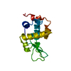
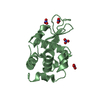
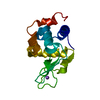
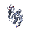
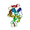
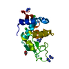
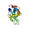
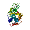
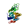
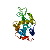
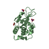
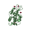
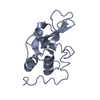
| Title | CRYSTAL STRUCTURE OF ORTHORHOMBIC LYSOZYME GROWN AT PH 6.5 AT 88% RELATIVE HUMIDITY | ||||||
 Components Components | LYSOZYME | ||||||
 Keywords Keywords | HYDROLASE / GLYCOSIDASE / ENZYME-ORTHORHOMBIC FORM / MUCOPEPTIDE N-ACETYLMURAMYL HYDROLASE / HEN EGG-WHITE LYSOZYME / LOW HUMIDITY | ||||||
| Function / homology |  Function and homology information Function and homology informationLactose synthesis / Antimicrobial peptides / Neutrophil degranulation / beta-N-acetylglucosaminidase activity / cell wall macromolecule catabolic process / lysozyme / lysozyme activity / killing of cells of another organism / defense response to Gram-negative bacterium / defense response to bacterium ...Lactose synthesis / Antimicrobial peptides / Neutrophil degranulation / beta-N-acetylglucosaminidase activity / cell wall macromolecule catabolic process / lysozyme / lysozyme activity / killing of cells of another organism / defense response to Gram-negative bacterium / defense response to bacterium / defense response to Gram-positive bacterium / endoplasmic reticulum / : / identical protein binding / cytoplasm Similarity search - Function | ||||||
| Biological species |  | ||||||
| Method |  X-RAY DIFFRACTION / Resolution: 1.7 Å X-RAY DIFFRACTION / Resolution: 1.7 Å | ||||||
 Authors Authors | Biswal, B.K. / Sukumar, N. / Vijayan, M. | ||||||
 Citation Citation | Journal: Acta Crystallogr.,Sect.D / Year: 2000 Title: Hydration, mobility and accessibility of lysozyme: structures of a pH 6.5 orthorhombic form and its low-humidity variant and a comparative study involving 20 crystallographically independent molecules. Authors: Biswal, B.K. / Sukumar, N. / Vijayan, M. #1: Journal: Acta Crystallogr.,Sect.D / Year: 1993Title: Protein Hydration and Water Structure: X-Ray Analysis of a Closely Packed Protein Crystal with Very Low Solvent Content Authors: Madhusudan / Kodandapani, R. / Vijayan, M. #2: Journal: Acta Crystallogr.,Sect.D / Year: 1999Title: Structures of Orthorhombic Lysozyme Grown at Basic pH and its Low-Humidity Variant Authors: Sukumar, N. / Biswal, B.K. / Vijayan, M. #3: Journal: J.Biol.Chem. / Year: 1990Title: Crystal Structure of Low Humidity Tetragonal Lysozyme at 2.1-A Resolution. Variability in Hydration Shell and its Structural Consequences Authors: Kodandapani, R. / Suresh, C.G. / Vijayan, M. | ||||||
| History |
|
- Structure visualization
| Structure viewer | Molecule: MolmilJmol/JSmol |
|---|
- Downloads & links
-Download
| PDBx/mmCIF format | 1f10.cif.gz | 39.9 KB | Display | PDBx/mmCIF format |
|---|---|---|---|---|
| PDB format | pdb1f10.ent.gz | 27.2 KB | Display | PDB format |
| PDBx/mmJSON format | 1f10.json.gz | Tree view | PDBx/mmJSON format | |
| Others |  Other downloads Other downloads |
-Validation report
| Arichive directory | https://data.pdbj.org/pub/pdb/validation_reports/f1/1f10ftp://data.pdbj.org/pub/pdb/validation_reports/f1/1f10 | HTTPS FTP |
|---|
-Related structure data


-Links
 PDBj
PDBj


- Assembly
| Deposited unit | 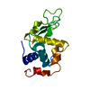
| ||||||||
|---|---|---|---|---|---|---|---|---|---|
| 1 |
| ||||||||
| Unit cell |
|
-Components
| #1: Protein | Mass: 14331.160 Da / Num. of mol.: 1 / Source method: isolated from a natural source / Source: (natural) |
|---|---|
| #2: Water | ChemComp-HOH /  Mass: 18.015 Da / Num. of mol.: 126 / Source method: isolated from a natural source / Formula: H2O Mass: 18.015 Da / Num. of mol.: 126 / Source method: isolated from a natural source / Formula: H2O |
| Has protein modification | Y |
-Experimental details
-Experiment
| Experiment | Method: X-RAY DIFFRACTION / Number of used crystals: 1 |
|---|
- Sample preparation
| Crystal | Density Matthews: 2.04 Å3/Da / Density % sol: 39.81 % | ||||||||||||||||||||||||||||||||||||
|---|---|---|---|---|---|---|---|---|---|---|---|---|---|---|---|---|---|---|---|---|---|---|---|---|---|---|---|---|---|---|---|---|---|---|---|---|---|
| Crystal grow | pH: 6.5 / Details: pH 6.5 | ||||||||||||||||||||||||||||||||||||
| Crystal | *PLUS Density % sol: 38.5 % | ||||||||||||||||||||||||||||||||||||
| Crystal grow | *PLUS Method: vapor diffusion, hanging drop | ||||||||||||||||||||||||||||||||||||
| Components of the solutions | *PLUS
|
-Data collection
| Diffraction | Mean temperature: 293 K |
|---|---|
| Diffraction source | Source: ROTATING ANODE / Type: RIGAKU RU200 / Wavelength: 1.5418 |
| Detector | Detector: IMAGE PLATE |
| Radiation | Protocol: SINGLE WAVELENGTH / Monochromatic (M) / Laue (L): M / Scattering type: x-ray |
| Radiation wavelength | Wavelength: 1.5418 Å / Relative weight: 1 |
| Reflection | Resolution: 1.7→25 Å / Num. obs: 13459 / % possible obs: 99.8 % / Redundancy: 4 % / Biso Wilson estimate: 18.9 Å2 / Rmerge(I) obs: 0.058 / Net I/σ(I): 25 |
| Reflection shell | Resolution: 1.7→1.8 Å / % possible obs: 99.9 % / Redundancy: 4.1 % / Rmerge(I) obs: 0.332 / Mean I/σ(I) obs: 3.1 / % possible all: 99.9 |
| Reflection | *PLUS Num. measured all: 54245 |
| Reflection shell | *PLUS Num. unique obs: 2083 |
- Processing
| Software |
| ||||||||||||||||||||||||||||||||||||
|---|---|---|---|---|---|---|---|---|---|---|---|---|---|---|---|---|---|---|---|---|---|---|---|---|---|---|---|---|---|---|---|---|---|---|---|---|---|
| Refinement | Starting model: ORTHORHOMBIC LYSOZYME AT PH 6.5 Resolution: 1.7→10 Å / Rfactor Rfree error: 0.008 / Data cutoff high absF: 10000000 / Data cutoff low absF: 0 / Isotropic thermal model: RESTRAINED / Cross valid method: THROUGHOUT / σ(F): 0
| ||||||||||||||||||||||||||||||||||||
| Displacement parameters | Biso mean: 20.8 Å2
| ||||||||||||||||||||||||||||||||||||
| Refine analyze |
| ||||||||||||||||||||||||||||||||||||
| Refinement step | Cycle: LAST / Resolution: 1.7→10 Å
| ||||||||||||||||||||||||||||||||||||
| Refine LS restraints |
| ||||||||||||||||||||||||||||||||||||
| LS refinement shell | Resolution: 1.7→1.81 Å / Rfactor Rfree error: 0.046 / Total num. of bins used: 6
| ||||||||||||||||||||||||||||||||||||
| Xplor file |
| ||||||||||||||||||||||||||||||||||||
| Software | *PLUS Name: X-PLOR / Version: 3.851 / Classification: refinement | ||||||||||||||||||||||||||||||||||||
| Refinement | *PLUS σ(F): 0 / % reflection Rfree: 10 % | ||||||||||||||||||||||||||||||||||||
| Solvent computation | *PLUS | ||||||||||||||||||||||||||||||||||||
| Displacement parameters | *PLUS Biso mean: 20.8 Å2 | ||||||||||||||||||||||||||||||||||||
| Refine LS restraints | *PLUS
| ||||||||||||||||||||||||||||||||||||
| LS refinement shell | *PLUS Rfactor Rfree: 0.486 / % reflection Rfree: 10.4 % / Rfactor Rwork: 0.414 |
