+ Open data
- Basic information
| Entry | Database: PDB / ID: 4dc4 | ||||||
|---|---|---|---|---|---|---|---|
| Title | Lysozyme Trimer | ||||||
 Components Components | Lysozyme C | ||||||
 Keywords Keywords | HYDROLASE | ||||||
| Function / homology |  Function and homology information Function and homology informationLactose synthesis / Antimicrobial peptides / Neutrophil degranulation / beta-N-acetylglucosaminidase activity / cell wall macromolecule catabolic process / lysozyme / lysozyme activity / killing of cells of another organism / defense response to Gram-negative bacterium / defense response to bacterium ...Lactose synthesis / Antimicrobial peptides / Neutrophil degranulation / beta-N-acetylglucosaminidase activity / cell wall macromolecule catabolic process / lysozyme / lysozyme activity / killing of cells of another organism / defense response to Gram-negative bacterium / defense response to bacterium / defense response to Gram-positive bacterium / endoplasmic reticulum / extracellular space / identical protein binding / cytoplasm Similarity search - Function | ||||||
| Biological species |  | ||||||
| Method |  X-RAY DIFFRACTION / X-RAY DIFFRACTION /  MOLECULAR REPLACEMENT / Resolution: 2.654 Å MOLECULAR REPLACEMENT / Resolution: 2.654 Å | ||||||
 Authors Authors | Sharma, P. / Ashish | ||||||
 Citation Citation |  Journal: Sci Rep / Year: 2016 Journal: Sci Rep / Year: 2016Title: Characterization of heat induced spherulites of lysozyme reveals new insight on amyloid initiation Authors: Sharma, P. / Verma, N. / Singh, P.K. / Korpole, S. / Ashish | ||||||
| History |
|
- Structure visualization
| Structure viewer | Molecule:  Molmil Molmil Jmol/JSmol Jmol/JSmol |
|---|
- Downloads & links
- Download
| PDBx/mmCIF format |  4dc4.cif.gz 4dc4.cif.gz | 86.9 KB | Display |  PDBx/mmCIF format PDBx/mmCIF format |
|---|---|---|---|---|
| PDB format |  pdb4dc4.ent.gz pdb4dc4.ent.gz | 66 KB | Display |  PDB format PDB format |
| PDBx/mmJSON format |  4dc4.json.gz 4dc4.json.gz | Tree view |  PDBx/mmJSON format PDBx/mmJSON format | |
| Others |  Other downloads Other downloads |
-Validation report
| Arichive directory |  https://data.pdbj.org/pub/pdb/validation_reports/dc/4dc4 https://data.pdbj.org/pub/pdb/validation_reports/dc/4dc4 ftp://data.pdbj.org/pub/pdb/validation_reports/dc/4dc4 ftp://data.pdbj.org/pub/pdb/validation_reports/dc/4dc4 | HTTPS FTP |
|---|
-Related structure data
| Related structure data |  4d9zC  4eofC 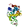 4ii8C 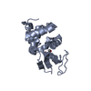 4r0fC 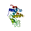 1bgiS S: Starting model for refinement C: citing same article ( |
|---|---|
| Similar structure data |
- Links
Links
- Assembly
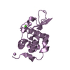
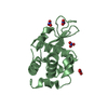
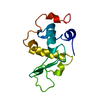
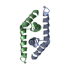
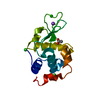
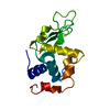
| Deposited unit | 
| ||||||||
|---|---|---|---|---|---|---|---|---|---|
| 1 | 
| ||||||||
| 2 | 
| ||||||||
| 3 | 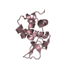
| ||||||||
| Unit cell |
| ||||||||
| Details | Author states that the biological assembly is eeakly associated lysozyme trimer |
- Components
| #1: Protein | Mass: 14331.160 Da / Num. of mol.: 3 / Source method: isolated from a natural source / Source: (natural)  #2: Chemical | #3: Chemical | #4: Chemical | #5: Water | ChemComp-HOH / | Has protein modification | Y | |
|---|
-Experimental details
-Experiment
| Experiment | Method:  X-RAY DIFFRACTION / Number of used crystals: 1 X-RAY DIFFRACTION / Number of used crystals: 1 |
|---|
- Sample preparation
| Crystal | Density Matthews: 1.93 Å3/Da / Density % sol: 36.29 % |
|---|---|
| Crystal grow | Temperature: 318 K / Method: vapor diffusion, hanging drop / pH: 3.8 Details: 50mM sodium acetate, 1-1.5M NaCl, pH 3.8, VAPOR DIFFUSION, HANGING DROP, temperature 318K |
-Data collection
| Diffraction | Mean temperature: 100 K |
|---|---|
| Diffraction source | Source:  ROTATING ANODE / Type: RIGAKU MICROMAX-007 HF / Wavelength: 1.5418 Å ROTATING ANODE / Type: RIGAKU MICROMAX-007 HF / Wavelength: 1.5418 Å |
| Detector | Type: MAR scanner 345 mm plate / Detector: IMAGE PLATE / Date: Dec 9, 2011 / Details: Mirrors |
| Radiation | Monochromator: Mirrors / Protocol: SINGLE WAVELENGTH / Monochromatic (M) / Laue (L): M / Scattering type: x-ray |
| Radiation wavelength | Wavelength: 1.5418 Å / Relative weight: 1 |
| Reflection | Resolution: 2.65→50 Å / Num. all: 10188 / Num. obs: 10188 / % possible obs: 92.9 % / Observed criterion σ(F): 2 / Observed criterion σ(I): 2 / Redundancy: 3.4 % / Biso Wilson estimate: 28.9 Å2 / Rmerge(I) obs: 0.188 / Net I/σ(I): 6.5 |
| Reflection shell | Resolution: 2.67→2.72 Å / Redundancy: 2 % / Rmerge(I) obs: 0.515 / Mean I/σ(I) obs: 2 / Num. unique all: 391 / % possible all: 82.9 |
- Processing
| Software |
| |||||||||||||||||||||||||
|---|---|---|---|---|---|---|---|---|---|---|---|---|---|---|---|---|---|---|---|---|---|---|---|---|---|---|
| Refinement | Method to determine structure:  MOLECULAR REPLACEMENT MOLECULAR REPLACEMENTStarting model: 1BGI Resolution: 2.654→35.909 Å / SU ML: 0.95 / Isotropic thermal model: Isotropic / σ(F): 0 / σ(I): 2 / Phase error: 29.93 / Stereochemistry target values: ML
| |||||||||||||||||||||||||
| Solvent computation | Shrinkage radii: 0.73 Å / VDW probe radii: 1 Å / Solvent model: FLAT BULK SOLVENT MODEL / Bsol: 29.606 Å2 / ksol: 0.41 e/Å3 | |||||||||||||||||||||||||
| Displacement parameters | Biso mean: 28.9 Å2
| |||||||||||||||||||||||||
| Refinement step | Cycle: LAST / Resolution: 2.654→35.909 Å
| |||||||||||||||||||||||||
| Refine LS restraints |
| |||||||||||||||||||||||||
| LS refinement shell | Refine-ID: X-RAY DIFFRACTION / Total num. of bins used: 3
|
 Movie
Movie Controller




 PDBj
PDBj


