 Movie
Movie Controller


+ Open data

- Basic information
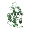
| Entry | Database: PDB / ID: 1hfx | ||||||
|---|---|---|---|---|---|---|---|
| Title | ALPHA-LACTALBUMIN | ||||||
 Components Components | ALPHA-LACTALBUMIN | ||||||
 Keywords Keywords | GLYCOPROTEIN / LACTOSE SYNTHASE COMPONENT / CALCIUM BINDING METALLOPROTEIN / LACTOSE | ||||||
| Function / homology |  Function and homology information Function and homology informationlactose synthase activity / lactose biosynthetic process / lysozyme activity / defense response to Gram-negative bacterium / defense response to Gram-positive bacterium / calcium ion binding / protein-containing complex / extracellular region Similarity search - Function | ||||||
| Biological species |  Cavia porcellus (domestic guinea pig) Cavia porcellus (domestic guinea pig) | ||||||
| Method |  X-RAY DIFFRACTION / MOLECULAR REPLACEMENT / Resolution: 1.9 Å X-RAY DIFFRACTION / MOLECULAR REPLACEMENT / Resolution: 1.9 Å | ||||||
 Authors Authors | Pike, A.C.W. / Brew, K. / Acharya, K.R. | ||||||
 Citation Citation | Journal: Structure / Year: 1996 Title: Crystal structures of guinea-pig, goat and bovine alpha-lactalbumin highlight the enhanced conformational flexibility of regions that are significant for its action in lactose synthase. Authors: Pike, A.C. / Brew, K. / Acharya, K.R. #1: Journal: J.Mol.Biol. / Year: 1991Title: Crystal Structure of Human Alpha-Lactalbumin at 1.7 A Resolution Authors: Acharya, K.R. / Ren, J.S. / Stuart, D.I. / Phillips, D.C. / Fenna, R.E. #2: Journal: J.Mol.Biol. / Year: 1989Title: Refined Structure of Baboon Alpha-Lactalbumin at 1.7 A Resolution. Comparison with C-Type Lysozyme Authors: Acharya, K.R. / Stuart, D.I. / Walker, N.P. / Lewis, M. / Phillips, D.C. | ||||||
| History |
|
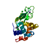
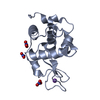
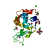
- Structure visualization
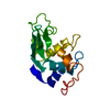
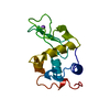
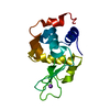
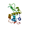
| Structure viewer | Molecule: MolmilJmol/JSmol |
|---|
- Downloads & links
-Download
| PDBx/mmCIF format | 1hfx.cif.gz | 38.7 KB | Display | PDBx/mmCIF format |
|---|---|---|---|---|
| PDB format | pdb1hfx.ent.gz | 25.9 KB | Display | PDB format |
| PDBx/mmJSON format | 1hfx.json.gz | Tree view | PDBx/mmJSON format | |
| Others |  Other downloads Other downloads |
-Validation report
| Arichive directory | https://data.pdbj.org/pub/pdb/validation_reports/hf/1hfxftp://data.pdbj.org/pub/pdb/validation_reports/hf/1hfx | HTTPS FTP |
|---|
-Related structure data

-Links
 PDBj
PDBj- Assembly
| Deposited unit | 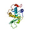
| ||||||||
|---|---|---|---|---|---|---|---|---|---|
| 1 |
| ||||||||
| Unit cell |
|
-Components
| #1: Protein | Mass: 14240.159 Da / Num. of mol.: 1 / Source method: isolated from a natural source / Source: (natural) Cavia porcellus (domestic guinea pig) / Secretion: MILK / References: UniProt: P00713, lactose synthase |
|---|---|
| #2: Chemical | ChemComp-CA /   Mass: 40.078 Da / Num. of mol.: 1 / Source method: obtained synthetically / Formula: Ca Mass: 40.078 Da / Num. of mol.: 1 / Source method: obtained synthetically / Formula: Ca |
| #3: Water | ChemComp-HOH /  Mass: 18.015 Da / Num. of mol.: 73 / Source method: isolated from a natural source / Formula: H2O Mass: 18.015 Da / Num. of mol.: 73 / Source method: isolated from a natural source / Formula: H2O |
| Has protein modification | Y |
-Experimental details
-Experiment
| Experiment | Method: X-RAY DIFFRACTION / Number of used crystals: 2 |
|---|
- Sample preparation
| Crystal | Density Matthews: 1.82 Å3/Da / Density % sol: 32 % | ||||||||||||||||||||||||||||||
|---|---|---|---|---|---|---|---|---|---|---|---|---|---|---|---|---|---|---|---|---|---|---|---|---|---|---|---|---|---|---|---|
| Crystal grow | Temperature: 293 K / pH: 4.6 Details: 0.2M AMMONIUM SULFATE, 20-25% (W/V) PEG 4000, 0.1M SODIUM ACETATE PH4.6, 20MG/ML PROTEIN 20 DEGREES C, temperature 293K | ||||||||||||||||||||||||||||||
| Crystal grow | *PLUS Temperature: 20 ℃ / Method: vapor diffusion, hanging drop | ||||||||||||||||||||||||||||||
| Components of the solutions | *PLUS
|
-Data collection
| Diffraction | Mean temperature: 298 K |
|---|---|
| Diffraction source | Wavelength: 1.5418 |
| Detector | Type: SIEMENS / Date: Feb 1, 1992 |
| Radiation | Monochromatic (M) / Laue (L): M / Scattering type: x-ray |
| Radiation wavelength | Wavelength: 1.5418 Å / Relative weight: 1 |
| Reflection | Resolution: 1.9→39.1 Å / Num. obs: 8003 / % possible obs: 88 % / Observed criterion σ(I): 0 / Redundancy: 6 % / Rmerge(I) obs: 0.083 |
- Processing
| Software |
| ||||||||||||||||||||||||||||||||||||||||||||||||||||||||||||
|---|---|---|---|---|---|---|---|---|---|---|---|---|---|---|---|---|---|---|---|---|---|---|---|---|---|---|---|---|---|---|---|---|---|---|---|---|---|---|---|---|---|---|---|---|---|---|---|---|---|---|---|---|---|---|---|---|---|---|---|---|---|
| Refinement | Method to determine structure: MOLECULAR REPLACEMENT Starting model: HUMAN ALPHA-LACTALBUMIN Resolution: 1.9→8 Å / σ(F): 0 /
| ||||||||||||||||||||||||||||||||||||||||||||||||||||||||||||
| Displacement parameters | Biso mean: 20.08 Å2 | ||||||||||||||||||||||||||||||||||||||||||||||||||||||||||||
| Refine analyze | Luzzati d res low obs: 5 Å / Luzzati sigma a obs: 0.19 Å | ||||||||||||||||||||||||||||||||||||||||||||||||||||||||||||
| Refinement step | Cycle: LAST / Resolution: 1.9→8 Å
| ||||||||||||||||||||||||||||||||||||||||||||||||||||||||||||
| Refine LS restraints |
| ||||||||||||||||||||||||||||||||||||||||||||||||||||||||||||
| Xplor file |
| ||||||||||||||||||||||||||||||||||||||||||||||||||||||||||||
| Software | *PLUS Name: X-PLOR / Version: 3.1 / Classification: refinement | ||||||||||||||||||||||||||||||||||||||||||||||||||||||||||||
| Refine LS restraints | *PLUS
|
