 Movie
Movie Controller

[English] 日本語
 Yorodumi
- PDB-1b2k: Structural effects of monovalent anions on polymorphic lysozyme c... -
+ Open data

- Basic information
| Entry | Database: PDB / ID: 1b2k | ||||||
|---|---|---|---|---|---|---|---|
| Title | Structural effects of monovalent anions on polymorphic lysozyme crystals | ||||||
 Components Components | PROTEIN (LYSOZYME) | ||||||
 Keywords Keywords | HYDROLASE / HYDROLASE (O-GLYCOSYL) | ||||||
| Function / homology |  Function and homology information Function and homology informationLactose synthesis / Antimicrobial peptides / Neutrophil degranulation / beta-N-acetylglucosaminidase activity / cell wall macromolecule catabolic process / lysozyme / lysozyme activity / killing of cells of another organism / defense response to Gram-negative bacterium / defense response to bacterium ...Lactose synthesis / Antimicrobial peptides / Neutrophil degranulation / beta-N-acetylglucosaminidase activity / cell wall macromolecule catabolic process / lysozyme / lysozyme activity / killing of cells of another organism / defense response to Gram-negative bacterium / defense response to bacterium / defense response to Gram-positive bacterium / endoplasmic reticulum / : / identical protein binding / cytoplasm Similarity search - Function | ||||||
| Biological species |  | ||||||
| Method |  X-RAY DIFFRACTION / SYNCHROTRON / MOLECULAR REPLACEMENT / Resolution: 1.6 Å X-RAY DIFFRACTION / SYNCHROTRON / MOLECULAR REPLACEMENT / Resolution: 1.6 Å | ||||||
 Authors Authors | Vaney, M.C. / Broutin, I. / Ries-Kautt, M. / Ducruix, A. | ||||||
 Citation Citation | Journal: Acta Crystallogr.,Sect.D / Year: 2001 Title: Structural effects of monovalent anions on polymorphic lysozyme crystals. Authors: Vaney, M.C. / Broutin, I. / Retailleau, P. / Douangamath, A. / Lafont, S. / Hamiaux, C. / Prange, T. / Ducruix, A. / Ries-Kautt, M. #1: Journal: Acta Crystallogr.,Sect.D / Year: 1998Title: Refinement of Triclinic Hen Egg-White Lysozyme at Atomic Resolution Authors: Walsh, M.A. / Schneider, T.R. / Sieker, L.C. / Dauter, Z. / Lamzin, V.S. / Wilson, K.S. #2: Journal: Acta Crystallogr.,Sect.D / Year: 1998Title: Structures of Monoclinic Lysozyme Iodide at 1.6 Ang. And of Triclinic Lysozyme Nitrate at 1.1 Ang Authors: Steinrauf, L.K. #3: Journal: Acta Crystallogr.,Sect.D / Year: 1998Title: Locations of Bromide Ions in Tetragonal Lysozyme Crystals Authors: Lim, K. / Nadarajah, A. / Forsythe, E.L. / Pusey, M.L. #4: Journal: Acta Crystallogr.,Sect.D / Year: 1996Title: High-Resolution Structure (1.33 Ang.) Of a Hewl Lysozyme Tetragonal Crystal Grown in the Apcf Apparatus. Data and Structural Comparison with a Crystal Grown Under Microgravity from Spachab-01 Mission Authors: Vaney, M.C. / Maignan, S. / Ries-Kautt, M. / Ducruix, A. | ||||||
| History |
|
- Structure visualization
| Structure viewer | Molecule: MolmilJmol/JSmol |
|---|
- Downloads & links
-Download
| PDBx/mmCIF format | 1b2k.cif.gz | 69.5 KB | Display | PDBx/mmCIF format |
|---|---|---|---|---|
| PDB format | pdb1b2k.ent.gz | 51.1 KB | Display | PDB format |
| PDBx/mmJSON format | 1b2k.json.gz | Tree view | PDBx/mmJSON format | |
| Others |  Other downloads Other downloads |
-Validation report
| Arichive directory | https://data.pdbj.org/pub/pdb/validation_reports/b2/1b2kftp://data.pdbj.org/pub/pdb/validation_reports/b2/1b2k | HTTPS FTP |
|---|
-Related structure data
| Related structure data | 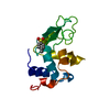 1b0dC 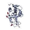 1hf4C 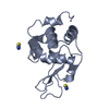 1lcnC 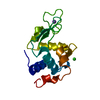 193lS S: Starting model for refinement C: citing same article ( |
|---|---|
| Similar structure data |


-Links
 PDBj
PDBj


- Assembly
| Deposited unit | 
| ||||||||
|---|---|---|---|---|---|---|---|---|---|
| 1 | 
| ||||||||
| 2 | 
| ||||||||
| Unit cell |
|
-Components
| #1: Protein | Mass: 14331.160 Da / Num. of mol.: 2 / Source method: isolated from a natural source / Source: (natural) #2: Chemical | ChemComp-IOD /   Mass: 126.904 Da / Num. of mol.: 17 / Source method: obtained synthetically / Formula: I Mass: 126.904 Da / Num. of mol.: 17 / Source method: obtained synthetically / Formula: I#3: Water | ChemComp-HOH / |  Mass: 18.015 Da / Num. of mol.: 291 / Source method: isolated from a natural source / Formula: H2O Mass: 18.015 Da / Num. of mol.: 291 / Source method: isolated from a natural source / Formula: H2OHas protein modification | Y | Nonpolymer details | WATER MOLECULE HOH 746, WELL-DEFINED IN THE DENSITY, IS HYDROGEN-BONDED WITH NE OF ARG 112A WITH A ...WATER MOLECULE HOH 746, WELL-DEFINED IN THE DENSITY, IS HYDROGEN-BONDED WITH NE OF ARG 112A WITH A DISTANCE OF 3.45 ANG. | |
|---|
-Experimental details
-Experiment
| Experiment | Method: X-RAY DIFFRACTION / Number of used crystals: 1 |
|---|
- Sample preparation
| Crystal | Density Matthews: 1.8 Å3/Da / Density % sol: 33 % | ||||||||||||||||||
|---|---|---|---|---|---|---|---|---|---|---|---|---|---|---|---|---|---|---|---|
| Crystal grow | Temperature: 291 K / pH: 4.5 Details: THE ISOIONIC PROTEIN HEWL WAS ACIDIFIED BY ADDING OF HI UNTIL THE PH 4.5 WAS REACHED AT 291K WERE 25 MG/ML OF PROTEIN AND 0.07 M OF NAI VERSUS A WELL CONTAINING 0.14 M OF NAI AT PH4.5 | ||||||||||||||||||
| Crystal grow | *PLUS Temperature: 291 K / Method: vapor diffusion, hanging dropDetails: pH is adjusted to 4.5 with HI, drop consists of equal volume of protein and reservoir solutions | ||||||||||||||||||
| Components of the solutions | *PLUS
|
-Data collection
| Diffraction | Mean temperature: 293 K |
|---|---|
| Diffraction source | Source: SYNCHROTRON / Site: LURE  / Beamline: DW32 / Wavelength: 0.901 / Beamline: DW32 / Wavelength: 0.901 |
| Detector | Type: MARRESEARCH / Detector: IMAGE PLATE / Date: Dec 1, 1992 |
| Radiation | Protocol: SINGLE WAVELENGTH / Monochromatic (M) / Laue (L): M / Scattering type: x-ray |
| Radiation wavelength | Wavelength: 0.901 Å / Relative weight: 1 |
| Reflection | Resolution: 1.6→13.3 Å / Num. obs: 26152 / % possible obs: 96.7 % / Observed criterion σ(I): 0 / Redundancy: 3.1 % / Biso Wilson estimate: 15.2 Å2 / Rsym value: 0.095 / Net I/σ(I): 5.2 |
| Reflection shell | Resolution: 1.6→1.64 Å / Redundancy: 2.8 % / Mean I/σ(I) obs: 1.3 / Rsym value: 0.54 / % possible all: 95.6 |
| Reflection | *PLUS Rmerge(I) obs: 0.095 |
| Reflection shell | *PLUS % possible obs: 95.6 % / Rmerge(I) obs: 0.5 |
- Processing
| Software |
| ||||||||||||||||||||||||||||||||||||||||||||||||||||||||||||||||||||||||||||||||
|---|---|---|---|---|---|---|---|---|---|---|---|---|---|---|---|---|---|---|---|---|---|---|---|---|---|---|---|---|---|---|---|---|---|---|---|---|---|---|---|---|---|---|---|---|---|---|---|---|---|---|---|---|---|---|---|---|---|---|---|---|---|---|---|---|---|---|---|---|---|---|---|---|---|---|---|---|---|---|---|---|---|
| Refinement | Method to determine structure: MOLECULAR REPLACEMENT Starting model: 193L Resolution: 1.6→8 Å / Data cutoff high absF: 10000000 / Data cutoff low absF: 0.001 / Isotropic thermal model: RESTRAINED / Cross valid method: THROUGHOUT / σ(F): 0
| ||||||||||||||||||||||||||||||||||||||||||||||||||||||||||||||||||||||||||||||||
| Displacement parameters | Biso mean: 16.4 Å2 | ||||||||||||||||||||||||||||||||||||||||||||||||||||||||||||||||||||||||||||||||
| Refine analyze |
| ||||||||||||||||||||||||||||||||||||||||||||||||||||||||||||||||||||||||||||||||
| Refinement step | Cycle: LAST / Resolution: 1.6→8 Å
| ||||||||||||||||||||||||||||||||||||||||||||||||||||||||||||||||||||||||||||||||
| Refine LS restraints |
| ||||||||||||||||||||||||||||||||||||||||||||||||||||||||||||||||||||||||||||||||
| LS refinement shell | Resolution: 1.6→1.67 Å / Total num. of bins used: 8
| ||||||||||||||||||||||||||||||||||||||||||||||||||||||||||||||||||||||||||||||||
| Xplor file |
| ||||||||||||||||||||||||||||||||||||||||||||||||||||||||||||||||||||||||||||||||
| Software | *PLUS Name: X-PLOR / Version: 3.851 / Classification: refinement | ||||||||||||||||||||||||||||||||||||||||||||||||||||||||||||||||||||||||||||||||
| Refine LS restraints | *PLUS
|
