Journal: Structure / Year: 2018 Title: A Rare Lysozyme Crystal Form Solved Using Highly Redundant Multiple Electron Diffraction Datasets from Micron-Sized Crystals. Authors: Hongyi Xu / Hugo Lebrette / Taimin Yang / Vivek Srinivas / Sven Hovmöller / Martin Högbom / Xiaodong Zou / Abstract: Recent developments of novel electron diffraction techniques have shown to be powerful for determination of atomic resolution structures from micron- and nano-sized crystals, too small to be studied ...Recent developments of novel electron diffraction techniques have shown to be powerful for determination of atomic resolution structures from micron- and nano-sized crystals, too small to be studied by single-crystal X-ray diffraction. In this work, the structure of a rare lysozyme polymorph is solved and refined using continuous rotation MicroED data and standard X-ray crystallographic software. Data collection was performed on a standard 200 kV transmission electron microscope (TEM) using a highly sensitive detector with a short readout time. The data collection is fast (∼3 min per crystal), allowing multiple datasets to be rapidly collected from a large number of crystals. We show that merging data from 33 crystals significantly improves not only the data completeness, overall I/σ and the data redundancy, but also the quality of the final atomic model. This is extremely useful for electron beam-sensitive crystals of low symmetry or with a preferred orientation on the TEM grid.
Mass: 18.015 Da / Num. of mol.: 25 / Source method: isolated from a natural source / Formula: H2O
Has protein modification
Y
-
Experimental details
-
Experiment
Experiment
Method: ELECTRON CRYSTALLOGRAPHY / Number of used crystals: 33
EM experiment
Aggregation state: 3D ARRAY / 3D reconstruction method: electron crystallography
-
Sample preparation
Component
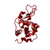
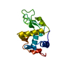
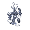
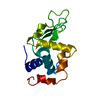
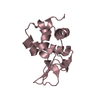
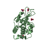
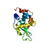
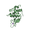
Name: 3D crystals of Lysozyme C from Gallus gallus / Type: COMPLEX / Entity ID: #1 / Source: NATURAL
Molecular weight
Experimental value: NO
Source (natural)
Organism: Gallus gallus (chicken)
EM crystal formation
Atmosphere: AIR Details: 2 microliters of Lysozyme (8 mg/ml in 10 mM Tris-HCl pH 8.0) was mixed with 2 microliters of precipitant solution consisting of 1 M potassium nitrate, 0.1 M sodium acetate trihydrate pH 3.4. ...Details: 2 microliters of Lysozyme (8 mg/ml in 10 mM Tris-HCl pH 8.0) was mixed with 2 microliters of precipitant solution consisting of 1 M potassium nitrate, 0.1 M sodium acetate trihydrate pH 3.4. Thin fibrous needle-shaped crystal clusters were grown by the hanging drop vapor diffusion method. Temperature: 294 K / Time: 2 DAY
Buffer solution
pH: 3.4
Buffer component
ID
Conc.
Name
Formula
Buffer-ID
1
1M
potassiumnitrate
KNO3
1
2
0.1M
sodiumacetate
C2H3NaO2
1
Specimen
Embedding applied: NO / Shadowing applied: NO / Staining applied: NO / Vitrification applied: YES
 Movie
Movie Controller

 Yorodumi
 Open data
 Basic information
 Components
 Keywords
 Function and homology information
 MOLECULAR REPLACEMENT / cryo EM / Resolution: 2.2 Å
  Authors
 Sweden, 6items
  Citation
 Structure visualization
 Downloads & links
 Other downloads
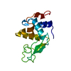


 PDBj
PDBj


 Assembly


 Mass: 22.990 Da / Num. of mol.: 1 / Source method: obtained synthetically / Formula: Na / Feature type: SUBJECT OF INVESTIGATION
 Mass: 18.015 Da / Num. of mol.: 25 / Source method: isolated from a natural source / Formula: H2O
 Sample preparation
 Processing