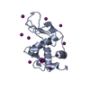
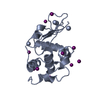
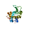
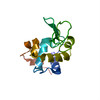
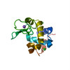
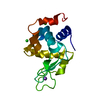
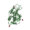
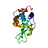
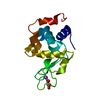
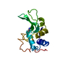
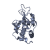
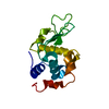
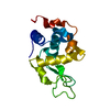
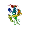
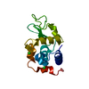
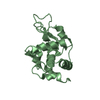
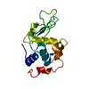
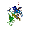
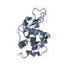
Journal: IUCrJ / Year: 2019 Title: Nanobeam precession-assisted 3D electron diffraction reveals a new polymorph of hen egg-white lysozyme. Authors: Arianna Lanza / Eleonora Margheritis / Enrico Mugnaioli / Valentina Cappello / Gianpiero Garau / Mauro Gemmi / Abstract: Recent advances in 3D electron diffraction have allowed the structure determination of several model proteins from submicrometric crystals, the unit-cell parameters and structures of which could be ...Recent advances in 3D electron diffraction have allowed the structure determination of several model proteins from submicrometric crystals, the unit-cell parameters and structures of which could be immediately validated by known models previously obtained by X-ray crystallography. Here, the first new protein structure determined by 3D electron diffraction data is presented: a previously unobserved polymorph of hen egg-white lysozyme. This form, with unit-cell parameters = 31.9, = 54.4, = 71.8 Å, β = 98.8°, grows as needle-shaped submicrometric crystals simply by vapor diffusion starting from previously reported crystallization conditions. Remarkably, the data were collected using a low-dose stepwise experimental setup consisting of a precession-assisted nanobeam of ∼150 nm, which has never previously been applied for solving protein structures. The crystal structure was additionally validated using X-ray synchrotron-radiation sources by both powder diffraction and single-crystal micro-diffraction. 3D electron diffraction can be used for the structural characterization of submicrometric macromolecular crystals and is able to identify novel protein polymorphs that are hardly visible in conventional X-ray diffraction experiments. Additionally, the analysis, which was performed on both nanocrystals and microcrystals from the same crystallization drop, suggests that an integrated view from 3D electron diffraction and X-ray microfocus diffraction can be applied to obtain insights into the molecular dynamics during protein crystal growth.
In the structure databanks used in Yorodumi, some data are registered as the other names, "COVID-19 virus" and "2019-nCoV". Here are the details of the virus and the list of structure data.
Jan 31, 2019. EMDB accession codes are about to change! (news from PDBe EMDB page)
EMDB accession codes are about to change! (news from PDBe EMDB page)
The allocation of 4 digits for EMDB accession codes will soon come to an end. Whilst these codes will remain in use, new EMDB accession codes will include an additional digit and will expand incrementally as the available range of codes is exhausted. The current 4-digit format prefixed with “EMD-” (i.e. EMD-XXXX) will advance to a 5-digit format (i.e. EMD-XXXXX), and so on. It is currently estimated that the 4-digit codes will be depleted around Spring 2019, at which point the 5-digit format will come into force.
The EM Navigator/Yorodumi systems omit the EMD- prefix.
Related info.:Q: What is EMD? / ID/Accession-code notation in Yorodumi/EM Navigator
Yorodumi is a browser for structure data from EMDB, PDB, SASBDB, etc.
This page is also the successor to EM Navigator detail page, and also detail information page/front-end page for Omokage search.
The word "yorodu" (or yorozu) is an old Japanese word meaning "ten thousand". "mi" (miru) is to see.
Related info.:EMDB / PDB / SASBDB / Comparison of 3 databanks / Yorodumi Search / Aug 31, 2016. New EM Navigator & Yorodumi / Yorodumi Papers / Jmol/JSmol / Function and homology information / Changes in new EM Navigator and Yorodumi
 Movie
Movie Controller

 Yorodumi
 Open data
 Basic information
 Components
 Keywords
 Function and homology information
 Authors
 Citation
 Structure visualization
 Downloads & links
 Other downloads
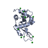


 PDBj
PDBj


 Assembly


 Mass: 35.453 Da / Num. of mol.: 3 / Source method: obtained synthetically / Formula: Cl
 Sample preparation
 Processing
 CCP4
CCP4