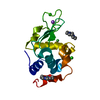
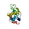
 Movie
Movie Controller

[English] 日本語
 Yorodumi
- PDB-5o4w: Protein structure determination by electron diffraction using a s... -
+ Open data

- Basic information
| Entry | Database: PDB / ID: 5o4w | ||||||
|---|---|---|---|---|---|---|---|
| Title | Protein structure determination by electron diffraction using a single three-dimensional nanocrystal | ||||||
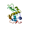
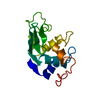
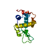
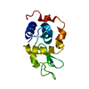
 Components Components | Lysozyme C | ||||||
 Keywords Keywords | HYDROLASE / Lysozyme / Nanocrystal | ||||||
| Function / homology |  Function and homology information Function and homology informationLactose synthesis / Antimicrobial peptides / Neutrophil degranulation / beta-N-acetylglucosaminidase activity / cell wall macromolecule catabolic process / lysozyme / lysozyme activity / killing of cells of another organism / defense response to Gram-negative bacterium / defense response to bacterium ...Lactose synthesis / Antimicrobial peptides / Neutrophil degranulation / beta-N-acetylglucosaminidase activity / cell wall macromolecule catabolic process / lysozyme / lysozyme activity / killing of cells of another organism / defense response to Gram-negative bacterium / defense response to bacterium / defense response to Gram-positive bacterium / endoplasmic reticulum / : / identical protein binding / cytoplasm Similarity search - Function | ||||||
| Biological species |  | ||||||
| Method | ELECTRON CRYSTALLOGRAPHY / electron crystallography /  MOLECULAR REPLACEMENT / cryo EM / Resolution: 2.11 Å MOLECULAR REPLACEMENT / cryo EM / Resolution: 2.11 Å | ||||||
 Authors Authors | Clabbers, M.T.B. / van Genderen, E. / Wan, W. / Wiegers, E.L. / Gruene, T. / Abrahams, J.P. | ||||||
 Citation Citation | Journal: Acta Crystallogr D Struct Biol / Year: 2017 Title: Protein structure determination by electron diffraction using a single three-dimensional nanocrystal. Authors: M T B Clabbers / E van Genderen / W Wan / E L Wiegers / T Gruene / J P Abrahams /    Abstract: Three-dimensional nanometre-sized crystals of macromolecules currently resist structure elucidation by single-crystal X-ray crystallography. Here, a single nanocrystal with a diffracting volume of ...Three-dimensional nanometre-sized crystals of macromolecules currently resist structure elucidation by single-crystal X-ray crystallography. Here, a single nanocrystal with a diffracting volume of only 0.14 µm, i.e. no more than 6 × 10 unit cells, provided sufficient information to determine the structure of a rare dimeric polymorph of hen egg-white lysozyme by electron crystallography. This is at least an order of magnitude smaller than was previously possible. The molecular-replacement solution, based on a monomeric polyalanine model, provided sufficient phasing power to show side-chain density, and automated model building was used to reconstruct the side chains. Diffraction data were acquired using the rotation method with parallel beam diffraction on a Titan Krios transmission electron microscope equipped with a novel in-house-designed 1024 × 1024 pixel Timepix hybrid pixel detector for low-dose diffraction data collection. Favourable detector characteristics include the ability to accurately discriminate single high-energy electrons from X-rays and count them, fast readout to finely sample reciprocal space and a high dynamic range. This work, together with other recent milestones, suggests that electron crystallography can provide an attractive alternative in determining biological structures. | ||||||
| History |
|
- Structure visualization
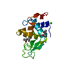
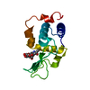
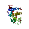
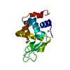
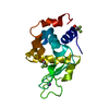
| Movie |
Movie viewer |
|---|---|
| Structure viewer | Molecule: MolmilJmol/JSmol |
- Downloads & links
-Download
| PDBx/mmCIF format | 5o4w.cif.gz | 61.6 KB | Display | PDBx/mmCIF format |
|---|---|---|---|---|
| PDB format | pdb5o4w.ent.gz | 44.3 KB | Display | PDB format |
| PDBx/mmJSON format | 5o4w.json.gz | Tree view | PDBx/mmJSON format | |
| Others |  Other downloads Other downloads |
-Validation report
| Arichive directory | https://data.pdbj.org/pub/pdb/validation_reports/o4/5o4wftp://data.pdbj.org/pub/pdb/validation_reports/o4/5o4w | HTTPS FTP |
|---|
-Related structure data
| Related structure data |  5o4xC 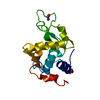 2yblS S: Starting model for refinement C: citing same article ( |
|---|---|
| Similar structure data |


-Links
 PDBj
PDBj


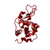
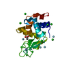
- Assembly
| Deposited unit | 
| ||||||||||||||||||||||||||||||
|---|---|---|---|---|---|---|---|---|---|---|---|---|---|---|---|---|---|---|---|---|---|---|---|---|---|---|---|---|---|---|---|
| 1 | 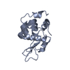
| ||||||||||||||||||||||||||||||
| 2 | 
| ||||||||||||||||||||||||||||||
| Unit cell |
| ||||||||||||||||||||||||||||||
| Noncrystallographic symmetry (NCS) | NCS domain:
NCS domain segments: Component-ID: 1 / Ens-ID: 1 / Beg auth comp-ID: LYS / Beg label comp-ID: LYS / End auth comp-ID: LEU / End label comp-ID: LEU / Refine code: 1 / Auth seq-ID: 1 - 129 / Label seq-ID: 1 - 129
NCS oper:
|
-Components
| #1: Protein | Mass: 14331.160 Da / Num. of mol.: 2 / Source method: isolated from a natural source / Source: (natural) Has protein modification | Y | |
|---|
-Experimental details
-Experiment
| Experiment | Method: ELECTRON CRYSTALLOGRAPHY |
|---|---|
| EM experiment | Aggregation state: 3D ARRAY / 3D reconstruction method: electron crystallography |
- Sample preparation
| Component | Name: Lysozyme C / Type: COMPLEX / Entity ID: all / Source: NATURAL |
|---|---|
| Source (natural) | Organism: |
| Buffer solution | pH: 3.8 |
| Specimen | Embedding applied: NO / Shadowing applied: NO / Staining applied: NO / Vitrification applied: YES |
| Vitrification | Cryogen name: ETHANE |
-Data collection
| Experimental equipment |  Model: Talos Arctica / Image courtesy: FEI Company |
|---|---|
| Microscopy | Model: FEI TALOS ARCTICA |
| Electron gun | Electron source: FIELD EMISSION GUN / Accelerating voltage: 200 kV / Illumination mode: OTHER |
| Electron lens | Mode: DIFFRACTION |
| Image recording | Electron dose: 0.01 e/Å2 / Film or detector model: OTHER |
| EM diffraction | Camera length: 1890 mm |
| EM diffraction shell | Resolution: 41.44→2.11 Å / Fourier space coverage: 48.38 % / Multiplicity: 1.9 / Num. of structure factors: 6717 / Phase residual: 179.9 ° |
| EM diffraction stats | Details: Parameters where extreme values were added are not applicable to our experiment and can not be filled out because there is no experimental data, these include overall phase error, overall ...Details: Parameters where extreme values were added are not applicable to our experiment and can not be filled out because there is no experimental data, these include overall phase error, overall phase residual and phase error rejection Fourier space coverage: 48.38 % / High resolution: 2.11 Å / Num. of intensities measured: 12601 / Num. of structure factors: 6717 / Phase error: 179.9 ° / Phase residual: 179.9 ° / Phase error rejection criteria: 9999 / Rmerge: 26.3 / Rsym: 26.4 |
- Processing
| Software | Name: REFMAC / Version: 5.8.0158 / Classification: refinement | ||||||||||||||||||||||||||||||||||||||||||||||||||||||||||||||||||||||||||||||||||||||||||||||||||||||||||
|---|---|---|---|---|---|---|---|---|---|---|---|---|---|---|---|---|---|---|---|---|---|---|---|---|---|---|---|---|---|---|---|---|---|---|---|---|---|---|---|---|---|---|---|---|---|---|---|---|---|---|---|---|---|---|---|---|---|---|---|---|---|---|---|---|---|---|---|---|---|---|---|---|---|---|---|---|---|---|---|---|---|---|---|---|---|---|---|---|---|---|---|---|---|---|---|---|---|---|---|---|---|---|---|---|---|---|---|
| EM 3D crystal entity | ∠α: 90 ° / ∠β: 90 ° / ∠γ: 90 ° / A: 104.56 Å / B: 68.05 Å / C: 32.05 Å / Space group name: P21212 / Space group num: 18 | ||||||||||||||||||||||||||||||||||||||||||||||||||||||||||||||||||||||||||||||||||||||||||||||||||||||||||
| CTF correction | Type: NONE | ||||||||||||||||||||||||||||||||||||||||||||||||||||||||||||||||||||||||||||||||||||||||||||||||||||||||||
| 3D reconstruction | Resolution method: DIFFRACTION PATTERN/LAYERLINES / Symmetry type: 3D CRYSTAL | ||||||||||||||||||||||||||||||||||||||||||||||||||||||||||||||||||||||||||||||||||||||||||||||||||||||||||
| Atomic model building | Protocol: OTHER | ||||||||||||||||||||||||||||||||||||||||||||||||||||||||||||||||||||||||||||||||||||||||||||||||||||||||||
| Refinement | Method to determine structure: MOLECULAR REPLACEMENT Starting model: 2ybl Resolution: 2.11→41.46 Å / Cor.coef. Fo:Fc: 0.848 / SU B: 10.205 / SU ML: 0.275 / Cross valid method: NONE Details: We present Rcomplete instead of Rfree, calculating Rcomplete takes into account all reflections which is preferred when considering data sets with less than about 10,000 unique reflections ...Details: We present Rcomplete instead of Rfree, calculating Rcomplete takes into account all reflections which is preferred when considering data sets with less than about 10,000 unique reflections [Brunger (1997) Methods in Enzymology 277:366-396, Luebben & Gruene (2015) PNAS 112:8999-9003]. To calculate Rcomplete a 0.2% test set size was chosen i.e. 500 test sets were created. First all non-measured observations were removed from the reflection file, and the 500 test sets were assigned at random. The model was then refined independently each time omitting one of the different test sets, thus in total 500 iterations. Each time refinement was carried out until practically reaching convergence. Finally, the value for Rcomplete was calculated from the excluded data i.e. calculated from all reflections. Since all structure factors are used in turn this leads to a more robust calculation than Rfree.
| ||||||||||||||||||||||||||||||||||||||||||||||||||||||||||||||||||||||||||||||||||||||||||||||||||||||||||
| Solvent computation | Ion probe radii: 0.8 Å / Shrinkage radii: 0.8 Å / VDW probe radii: 1.2 Å | ||||||||||||||||||||||||||||||||||||||||||||||||||||||||||||||||||||||||||||||||||||||||||||||||||||||||||
| Displacement parameters | Biso mean: 23.986 Å2
| ||||||||||||||||||||||||||||||||||||||||||||||||||||||||||||||||||||||||||||||||||||||||||||||||||||||||||
| Refinement step | Cycle: 1 / Total: 2000 | ||||||||||||||||||||||||||||||||||||||||||||||||||||||||||||||||||||||||||||||||||||||||||||||||||||||||||
| Refine LS restraints |
|