 Movie
Movie Controller

[English] 日本語
 Yorodumi
- PDB-1bwi: THE 1.8 A STRUCTURE OF MICROBATCH OIL DROP GROWN TETRAGONAL HEN E... -
+ Open data

- Basic information
| Entry | Database: PDB / ID: 1bwi | ||||||
|---|---|---|---|---|---|---|---|
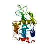
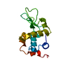
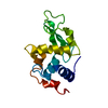
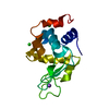
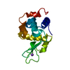
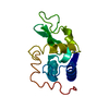
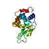
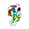
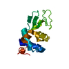
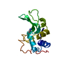
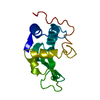
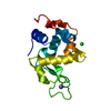
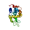
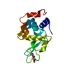
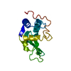
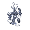
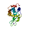
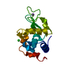
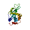
| Title | THE 1.8 A STRUCTURE OF MICROBATCH OIL DROP GROWN TETRAGONAL HEN EGG WHITE LYSOZYME | ||||||
 Components Components | PROTEIN (LYSOZYME) | ||||||
 Keywords Keywords | HYDROLASE / LYSOZYME / 1 / 4-BETA-N-ACETYLMURAMIDASE C | ||||||
| Function / homology |  Function and homology information Function and homology informationLactose synthesis / Antimicrobial peptides / Neutrophil degranulation / beta-N-acetylglucosaminidase activity / cell wall macromolecule catabolic process / lysozyme / lysozyme activity / killing of cells of another organism / defense response to Gram-negative bacterium / defense response to bacterium ...Lactose synthesis / Antimicrobial peptides / Neutrophil degranulation / beta-N-acetylglucosaminidase activity / cell wall macromolecule catabolic process / lysozyme / lysozyme activity / killing of cells of another organism / defense response to Gram-negative bacterium / defense response to bacterium / defense response to Gram-positive bacterium / endoplasmic reticulum / : / identical protein binding / cytoplasm Similarity search - Function | ||||||
| Biological species |  | ||||||
| Method |  X-RAY DIFFRACTION / MOLECULAR REPLACEMENT / Resolution: 1.8 Å X-RAY DIFFRACTION / MOLECULAR REPLACEMENT / Resolution: 1.8 Å | ||||||
 Authors Authors | Dong, J. / Boggon, T.J. / Chayen, N.E. / Raftery, J. / Bi, R.C. | ||||||
 Citation Citation | Journal: Acta Crystallogr.,Sect.D / Year: 1999 Title: Bound-solvent structures for microgravity-, ground control-, gel- and microbatch-grown hen egg-white lysozyme crystals at 1.8 A resolution. Authors: Dong, J. / Boggon, T.J. / Chayen, N.E. / Raftery, J. / Bi, R.C. / Helliwell, J.R. | ||||||
| History |
|
- Structure visualization
| Structure viewer | Molecule: MolmilJmol/JSmol |
|---|
- Downloads & links
-Download
| PDBx/mmCIF format | 1bwi.cif.gz | 38.5 KB | Display | PDBx/mmCIF format |
|---|---|---|---|---|
| PDB format | pdb1bwi.ent.gz | 26.2 KB | Display | PDB format |
| PDBx/mmJSON format | 1bwi.json.gz | Tree view | PDBx/mmJSON format | |
| Others |  Other downloads Other downloads |
-Validation report
| Arichive directory | https://data.pdbj.org/pub/pdb/validation_reports/bw/1bwiftp://data.pdbj.org/pub/pdb/validation_reports/bw/1bwi | HTTPS FTP |
|---|
-Related structure data
| Related structure data | 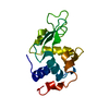 1bvxC 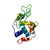 1bwhC 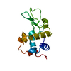 1bwjC 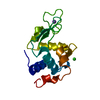 193lS S: Starting model for refinement C: citing same article ( |
|---|---|
| Similar structure data |

-Links
 PDBj
PDBj


- Assembly
| Deposited unit | 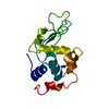
| ||||||||
|---|---|---|---|---|---|---|---|---|---|
| 1 |
| ||||||||
| Unit cell |
|
-Components
| #1: Protein | Mass: 14331.160 Da / Num. of mol.: 1 / Source method: isolated from a natural source / Details: SIGMA / Source: (natural) |
|---|---|
| #2: Water | ChemComp-HOH /  Mass: 18.015 Da / Num. of mol.: 92 / Source method: isolated from a natural source / Formula: H2O Mass: 18.015 Da / Num. of mol.: 92 / Source method: isolated from a natural source / Formula: H2O |
| Has protein modification | Y |
-Experimental details
-Experiment
| Experiment | Method: X-RAY DIFFRACTION / Number of used crystals: 1 |
|---|
- Sample preparation
| Crystal | Density Matthews: 2.08 Å3/Da / Density % sol: 40.78 % | ||||||||||||||||||||||||
|---|---|---|---|---|---|---|---|---|---|---|---|---|---|---|---|---|---|---|---|---|---|---|---|---|---|
| Crystal grow | Temperature: 291 K / Method: drop under paraffin oil / pH: 4.7 Details: A SOLUTION CONTAINING 40 MG/ML OF LYSOZYME (SIGMA L-6876, BATCH NUMBER, LOT 65H7025, IE A DIFFERENT SIGMA SAMPLE TO THE EXPERIMENTS DESCRIBED ABOVE) IN 10 MM SODIUM CITRATE BUFFER PH 4.7 WAS ...Details: A SOLUTION CONTAINING 40 MG/ML OF LYSOZYME (SIGMA L-6876, BATCH NUMBER, LOT 65H7025, IE A DIFFERENT SIGMA SAMPLE TO THE EXPERIMENTS DESCRIBED ABOVE) IN 10 MM SODIUM CITRATE BUFFER PH 4.7 WAS MIXED IN AN EPPENDORF TUBE WITH EQUAL VOLUMES OF 12 % SODIUM CHLORIDE (BDH). THE MIXTURE WAS FILTERED THROUGH A 0.22 UM FILTER AND 8 UL DROPS WERE MANUALLY DISPENSED UNDER PARAFFIN OIL USING A GILSON MICRO PIPPETTE. OVER 20 IDENTICAL DROPS WERE SET UP AND, WITHIN A WEEK, SINGLE CRYSTALS TYPICALLY MEASURING 0.5 X 0.4 X 0.3 MM WERE OBTAINED IN MOST OF THE DROPS. CRYSTALLISATION TOOK PLACE AT A TEMPERATURE OF 18 - 20 C., drops under paraffin oil, temperature 291K | ||||||||||||||||||||||||
| Crystal grow | *PLUS Method: microbatch method | ||||||||||||||||||||||||
| Components of the solutions | *PLUS
|
-Data collection
| Diffraction | Mean temperature: 293 K |
|---|---|
| Diffraction source | Source: ROTATING ANODE / Type: RIGAKU RU200 / Wavelength: 1.5418 |
| Detector | Type: RIGAKU RAXIS IIC / Detector: IMAGE PLATE |
| Radiation | Monochromator: CARBON / Protocol: SINGLE WAVELENGTH / Monochromatic (M) / Laue (L): M / Scattering type: x-ray |
| Radiation wavelength | Wavelength: 1.5418 Å / Relative weight: 1 |
| Reflection | Resolution: 1.7→99 Å / Num. obs: 12081 / % possible obs: 85.5 % / Redundancy: 6.1 % / Rmerge(I) obs: 0.077 / Rsym value: 0.08 / Net I/σ(I): 22.6 |
| Reflection | *PLUS % possible obs: 87.2 % / Num. measured all: 73591 |
| Reflection shell | *PLUS Highest resolution: 1.7 Å / Lowest resolution: 1.74 Å / % possible obs: 43.2 % / Rmerge(I) obs: 0.151 |
- Processing
| Software |
| ||||||||||||||||||||||||||||||||||||||||||||||||||||||||||||
|---|---|---|---|---|---|---|---|---|---|---|---|---|---|---|---|---|---|---|---|---|---|---|---|---|---|---|---|---|---|---|---|---|---|---|---|---|---|---|---|---|---|---|---|---|---|---|---|---|---|---|---|---|---|---|---|---|---|---|---|---|---|
| Refinement | Method to determine structure: MOLECULAR REPLACEMENT Starting model: PDB ENTRY 193L Resolution: 1.8→20 Å / Data cutoff high absF: 10000000 / Data cutoff low absF: 0.001 / Cross valid method: THROUGHOUT / σ(F): 0
| ||||||||||||||||||||||||||||||||||||||||||||||||||||||||||||
| Displacement parameters | Biso mean: 22.9 Å2 | ||||||||||||||||||||||||||||||||||||||||||||||||||||||||||||
| Refinement step | Cycle: LAST / Resolution: 1.8→20 Å
| ||||||||||||||||||||||||||||||||||||||||||||||||||||||||||||
| Refine LS restraints |
| ||||||||||||||||||||||||||||||||||||||||||||||||||||||||||||
| Software | *PLUS Name: X-PLOR / Version: 3.1 / Classification: refinement | ||||||||||||||||||||||||||||||||||||||||||||||||||||||||||||
| Refinement | *PLUS σ(F): 0 / % reflection Rfree: 5.1 % | ||||||||||||||||||||||||||||||||||||||||||||||||||||||||||||
| Solvent computation | *PLUS | ||||||||||||||||||||||||||||||||||||||||||||||||||||||||||||
| Displacement parameters | *PLUS Biso mean: 22.9 Å2 | ||||||||||||||||||||||||||||||||||||||||||||||||||||||||||||
| Refine LS restraints | *PLUS
| ||||||||||||||||||||||||||||||||||||||||||||||||||||||||||||
| LS refinement shell | *PLUS Highest resolution: 1.8 Å / Lowest resolution: 1.88 Å / Rfactor Rfree: 0.329 / Rfactor obs: 0.259 |
