[English] 日本語
Yorodumi
- PDB-5a3e: 2.5A structure of lysozyme determined by MicroED with data from a... -

+
Open data


ID or keywords:

Loading...

-
Basic information

Entry
Database: PDB / ID: 5a3e
Title2.5A structure of lysozyme determined by MicroED with data from a single crystal
ComponentsLYSOZYME C
KeywordsHYDROLASE / MICROED
Function / homology
Function and homology information


Antimicrobial peptides / Neutrophil degranulation / beta-N-acetylglucosaminidase activity / cell wall macromolecule catabolic process / lysozyme / lysozyme activity / killing of cells of another organism / defense response to Gram-negative bacterium / defense response to Gram-positive bacterium / defense response to bacterium ...Antimicrobial peptides / Neutrophil degranulation / beta-N-acetylglucosaminidase activity / cell wall macromolecule catabolic process / lysozyme / lysozyme activity / killing of cells of another organism / defense response to Gram-negative bacterium / defense response to Gram-positive bacterium / defense response to bacterium / endoplasmic reticulum / extracellular space / identical protein binding / cytoplasm
Similarity search - Function
Lysozyme - #10 / Glycoside hydrolase, family 22, lysozyme / Glycoside hydrolase family 22 domain / Glycosyl hydrolases family 22 (GH22) domain signature. / Glycoside hydrolase, family 22 / C-type lysozyme/alpha-lactalbumin family / Glycosyl hydrolases family 22 (GH22) domain profile. / Alpha-lactalbumin / lysozyme C / Lysozyme / Lysozyme-like domain superfamily ...Lysozyme - #10 / Glycoside hydrolase, family 22, lysozyme / Glycoside hydrolase family 22 domain / Glycosyl hydrolases family 22 (GH22) domain signature. / Glycoside hydrolase, family 22 / C-type lysozyme/alpha-lactalbumin family / Glycosyl hydrolases family 22 (GH22) domain profile. / Alpha-lactalbumin / lysozyme C / Lysozyme / Lysozyme-like domain superfamily / Orthogonal Bundle / Mainly Alpha
Similarity search - Domain/homology
Biological speciesGALLUS GALLUS (chicken)
MethodELECTRON CRYSTALLOGRAPHY / electron crystallography / 
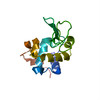
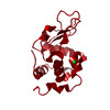
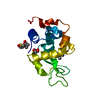
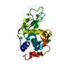
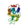
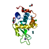
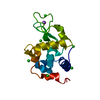
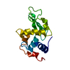
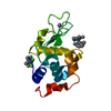
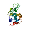
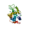
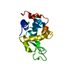
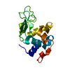
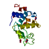
MOLECULAR REPLACEMENT / cryo EM / Resolution: 2.501 Å
AuthorsNannenga, B.L. / Shi, D. / Leslie, A.G.W. / Gonen, T.
CitationJournal: Nat Methods / Year: 2014
Title: High-resolution structure determination by continuous-rotation data collection in MicroED.
Authors: Brent L Nannenga / Dan Shi / Andrew G W Leslie / Tamir Gonen /
Abstract: MicroED uses very small three-dimensional protein crystals and electron diffraction for structure determination. We present an improved data collection protocol for MicroED called 'continuous ...MicroED uses very small three-dimensional protein crystals and electron diffraction for structure determination. We present an improved data collection protocol for MicroED called 'continuous rotation'. Microcrystals are continuously rotated during data collection, yielding more accurate data. The method enables data processing with the crystallographic software tool MOSFLM, which resulted in improved resolution for the model protein lysozyme. These improvements are paving the way for the broad implementation and application of MicroED in structural biology.
History
DepositionMay 28, 2015Deposition site: PDBE / Processing site: PDBE
Revision 1.0Jun 17, 2015Provider: repository / Type: Initial release
Revision 1.1Apr 19, 2017Group: Other
Revision 1.2Jan 10, 2024Group: Author supporting evidence / Data collection ...Author supporting evidence / Data collection / Database references / Refinement description
Category: chem_comp_atom / chem_comp_bond ...chem_comp_atom / chem_comp_bond / database_2 / diffrn_radiation / em_image_scans / em_single_particle_entity / pdbx_initial_refinement_model
Item: _database_2.pdbx_DOI / _database_2.pdbx_database_accession / _diffrn_radiation.pdbx_scattering_type

-
Structure visualization

Movie
  • Deposited structure unit
  • Imaged by Jmol
  • Download
  • Simplified surface model + fitted atomic model
  • EMDB-6342
  • Imaged by Jmol
  • Download
  • Superimposition on EM map
  • EMDB-6342
  • Imaged by UCSF Chimera
  • Download
Movie viewer
Structure viewerMolecule:
MolmilJmol/JSmol

Downloads & links

-
Assembly

Deposited unit
A: LYSOZYME C


Theoretical massNumber of molelcules
Total (without water)14,3311
Polymers14,3311
Non-polymers00
Water18010
1


  • Idetical with deposited unit
  • defined by author&software
TypeNameSymmetry operationNumber
identity operation1_555x,y,z1
MethodPISA
Unit cell
Length a, b, c (Å)75.890, 75.890, 36.930
Angle α, β, γ (deg.)90.00, 90.00, 90.00
Int Tables number96
Space group name H-MP43212

-
Components

#1: Protein LYSOZYME C / 1 / 4-BETA-N-ACETYLMURAMIDASE C / ALLERGEN GAL D IV


Mass: 14331.160 Da / Num. of mol.: 1 / Source method: isolated from a natural source / Details: EGG WHITES / Source: (natural) GALLUS GALLUS (chicken) / References: UniProt: P00698, lysozyme
#2: Water ChemComp-HOH / water / Water


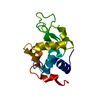
Mass: 18.015 Da / Num. of mol.: 10 / Source method: isolated from a natural source / Formula: H2O

-
Experimental details

-
Experiment

ExperimentMethod: ELECTRON CRYSTALLOGRAPHY / Number of used crystals: 1
EM experimentAggregation state: 3D ARRAY / 3D reconstruction method: electron crystallography

-
Sample preparation

ComponentName: Lysozyme / Type: COMPLEX
SpecimenEmbedding applied: NO / Shadowing applied: NO / Staining applied: NO / Vitrification applied: YES
Crystal growpH: 4.5 / Details: pH 4.5

-
Data collection

Experimental equipment
Model: Tecnai F20 / Image courtesy: FEI Company
MicroscopyModel: FEI TECNAI F20
Electron gunAccelerating voltage: 200 kV / Illumination mode: FLOOD BEAM
Electron lensMode: DIFFRACTION
Image recordingFilm or detector model: GENERIC CCD
DiffractionMean temperature: 100 K
DetectorDate: Feb 5, 2014
RadiationScattering type: electron
Radiation wavelengthRelative weight: 1
ReflectionResolution: 2.5→11.2 Å / Num. obs: 3116 / % possible obs: 80.1 % / Redundancy: 3.4 %
Reflection shellResolution: 2.5→2.6 Å / Redundancy: 3.2 % / Rmerge(I) obs: 0.292 / % possible all: 80.1

-
Processing

Software
NameVersionClassification
PHASERmodel building
PHENIX(PHENIX.REFINE)refinement
Aimlessdata scaling
PHASERphasing
3D reconstructionSymmetry type: 3D CRYSTAL
RefinementMethod to determine structure: MOLECULAR REPLACEMENT
Starting model: PDB ENTRY 3J6K
Resolution: 2.501→11.189 Å / SU ML: 0.23 / σ(F): 1.46 / Phase error: 21.2 / Stereochemistry target values: ML
RfactorNum. reflection% reflection
Rfree0.2531 142 4.6 %
Rwork0.2131 --
obs0.2152 3115 78.35 %
Solvent computationShrinkage radii: 0.9 Å / VDW probe radii: 1.11 Å / Solvent model: FLAT BULK SOLVENT MODEL
Refinement stepCycle: LAST / Resolution: 2.501→11.189 Å
ProteinNucleic acidLigandSolventTotal
Num. atoms992 0 0 10 1002
Refine LS restraints
Refine-IDTypeDev idealNumber
ELECTRON CRYSTALLOGRAPHYf_bond_d0.0031016
ELECTRON CRYSTALLOGRAPHYf_angle_d0.6871368
ELECTRON CRYSTALLOGRAPHYf_dihedral_angle_d12.347361
ELECTRON CRYSTALLOGRAPHYf_chiral_restr0.03142
ELECTRON CRYSTALLOGRAPHYf_plane_restr0.004179
LS refinement shellResolution: 2.5006→11.1887 Å
RfactorNum. reflection% reflection
Rfree0.2531 142 -
Rwork0.2131 2973 -
obs--78 %

+
About Yorodumi

-
News

-
Feb 9, 2022. New format data for meta-information of EMDB entries

New format data for meta-information of EMDB entries

  • Version 3 of the EMDB header file is now the official format.
  • The previous official version 1.9 will be removed from the archive.

Related info.:EMDB header

External links:wwPDB to switch to version 3 of the EMDB data model

-
Aug 12, 2020. Covid-19 info

Covid-19 info

URL: https://pdbj.org/emnavi/covid19.php

New page: Covid-19 featured information page in EM Navigator.

Related info.:Covid-19 info / Mar 5, 2020. Novel coronavirus structure data

+
Mar 5, 2020. Novel coronavirus structure data

Novel coronavirus structure data

Related info.:Yorodumi Speices / Aug 12, 2020. Covid-19 info

External links:COVID-19 featured content - PDBj / Molecule of the Month (242):Coronavirus Proteases

+
Jan 31, 2019. EMDB accession codes are about to change! (news from PDBe EMDB page)

EMDB accession codes are about to change! (news from PDBe EMDB page)

  • The allocation of 4 digits for EMDB accession codes will soon come to an end. Whilst these codes will remain in use, new EMDB accession codes will include an additional digit and will expand incrementally as the available range of codes is exhausted. The current 4-digit format prefixed with “EMD-” (i.e. EMD-XXXX) will advance to a 5-digit format (i.e. EMD-XXXXX), and so on. It is currently estimated that the 4-digit codes will be depleted around Spring 2019, at which point the 5-digit format will come into force.
  • The EM Navigator/Yorodumi systems omit the EMD- prefix.

Related info.:Q: What is EMD? / ID/Accession-code notation in Yorodumi/EM Navigator

External links:EMDB Accession Codes are Changing Soon! / Contact to PDBj

+
Jul 12, 2017. Major update of PDB

Major update of PDB

  • wwPDB released updated PDB data conforming to the new PDBx/mmCIF dictionary.
  • This is a major update changing the version number from 4 to 5, and with Remediation, in which all the entries are updated.
  • In this update, many items about electron microscopy experimental information are reorganized (e.g. em_software).
  • Now, EM Navigator and Yorodumi are based on the updated data.

External links:wwPDB Remediation / Enriched Model Files Conforming to OneDep Data Standards Now Available in the PDB FTP Archive

-
Yorodumi

Thousand views of thousand structures

  • Yorodumi is a browser for structure data from EMDB, PDB, SASBDB, etc.
  • This page is also the successor to EM Navigator detail page, and also detail information page/front-end page for Omokage search.
  • The word "yorodu" (or yorozu) is an old Japanese word meaning "ten thousand". "mi" (miru) is to see.

Related info.:EMDB / PDB / SASBDB / Comparison of 3 databanks / Yorodumi Search / Aug 31, 2016. New EM Navigator & Yorodumi / Yorodumi Papers / Jmol/JSmol / Function and homology information / Changes in new EM Navigator and Yorodumi

Read more