 Movie
Movie Controller

[English] 日本語
 Yorodumi
- PDB-5njm: Lysozyme room-temperature structure determined by serial millisec... -
+ Open data

- Basic information
| Entry | Database: PDB / ID: 5njm | ||||||
|---|---|---|---|---|---|---|---|
| Title | Lysozyme room-temperature structure determined by serial millisecond crystallography | ||||||
 Components Components | Lysozyme C | ||||||
 Keywords Keywords | HYDROLASE / room-temperature / serial crystallography | ||||||
| Function / homology |  Function and homology information Function and homology informationLactose synthesis / Antimicrobial peptides / Neutrophil degranulation / beta-N-acetylglucosaminidase activity / cell wall macromolecule catabolic process / lysozyme / lysozyme activity / killing of cells of another organism / defense response to Gram-negative bacterium / defense response to bacterium ...Lactose synthesis / Antimicrobial peptides / Neutrophil degranulation / beta-N-acetylglucosaminidase activity / cell wall macromolecule catabolic process / lysozyme / lysozyme activity / killing of cells of another organism / defense response to Gram-negative bacterium / defense response to bacterium / defense response to Gram-positive bacterium / endoplasmic reticulum / : / identical protein binding / cytoplasm Similarity search - Function | ||||||
| Biological species |  | ||||||
| Method |  X-RAY DIFFRACTION / SYNCHROTRON / MOLECULAR REPLACEMENT / Resolution: 1.5 Å X-RAY DIFFRACTION / SYNCHROTRON / MOLECULAR REPLACEMENT / Resolution: 1.5 Å | ||||||
 Authors Authors | Weinert, T. / Vera, L. / Marsh, M. / James, D. / Gashi, D. / Nogly, P. / Jaeger, K. / Standfuss, J. | ||||||
 Citation Citation | Journal: Nat Commun / Year: 2017 Title: Serial millisecond crystallography for routine room-temperature structure determination at synchrotrons. Authors: Weinert, T. / Olieric, N. / Cheng, R. / Brunle, S. / James, D. / Ozerov, D. / Gashi, D. / Vera, L. / Marsh, M. / Jaeger, K. / Dworkowski, F. / Panepucci, E. / Basu, S. / Skopintsev, P. / ...Authors: Weinert, T. / Olieric, N. / Cheng, R. / Brunle, S. / James, D. / Ozerov, D. / Gashi, D. / Vera, L. / Marsh, M. / Jaeger, K. / Dworkowski, F. / Panepucci, E. / Basu, S. / Skopintsev, P. / Dore, A.S. / Geng, T. / Cooke, R.M. / Liang, M. / Prota, A.E. / Panneels, V. / Nogly, P. / Ermler, U. / Schertler, G. / Hennig, M. / Steinmetz, M.O. / Wang, M. / Standfuss, J. | ||||||
| History |
|
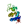
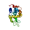
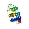
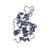
- Structure visualization
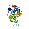
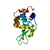
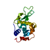
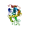
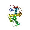
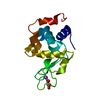
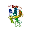
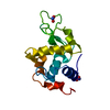
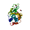
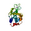
| Structure viewer | Molecule: MolmilJmol/JSmol |
|---|
- Downloads & links
-Download
| PDBx/mmCIF format | 5njm.cif.gz | 87 KB | Display | PDBx/mmCIF format |
|---|---|---|---|---|
| PDB format | pdb5njm.ent.gz | 66.3 KB | Display | PDB format |
| PDBx/mmJSON format | 5njm.json.gz | Tree view | PDBx/mmJSON format | |
| Others |  Other downloads Other downloads |
-Validation report
| Arichive directory | https://data.pdbj.org/pub/pdb/validation_reports/nj/5njmftp://data.pdbj.org/pub/pdb/validation_reports/nj/5njm | HTTPS FTP |
|---|
-Related structure data
| Related structure data | 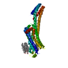 5nlxC 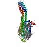 5nm2C 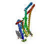 5nm4C 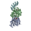 5nm5C 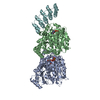 5nqtC 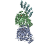 5nquC 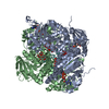 5o5wC 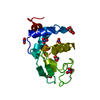 3lztS S: Starting model for refinement C: citing same article ( |
|---|---|
| Similar structure data |


-Links
 PDBj
PDBj


- Assembly
| Deposited unit | 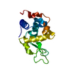
| ||||||||
|---|---|---|---|---|---|---|---|---|---|
| 1 |
| ||||||||
| Unit cell |
| ||||||||
| Components on special symmetry positions |
|
-Components
| #1: Protein | Mass: 14331.160 Da / Num. of mol.: 1 / Source method: isolated from a natural source / Source: (natural) |
|---|---|
| #2: Water | ChemComp-HOH /  Mass: 18.015 Da / Num. of mol.: 69 / Source method: isolated from a natural source / Formula: H2O Mass: 18.015 Da / Num. of mol.: 69 / Source method: isolated from a natural source / Formula: H2O |
| Has protein modification | Y |
-Experimental details
-Experiment
| Experiment | Method: X-RAY DIFFRACTION / Number of used crystals: 1 |
|---|
- Sample preparation
| Crystal | Density Matthews: 2.09 Å3/Da / Density % sol: 41.22 % |
|---|---|
| Crystal grow | Temperature: 293 K / Method: batch mode / pH: 3 Details: 19.04% NaCl, 5.44% PEG 8,000, 68mM Na Acetate pH 3.0 |
-Data collection
| Diffraction | Mean temperature: 293 K |
|---|---|
| Diffraction source | Source: SYNCHROTRON / Site: SLS  / Beamline: X06SA / Wavelength: 1 Å / Beamline: X06SA / Wavelength: 1 Å |
| Detector | Type: DECTRIS EIGER X 16M / Detector: PIXEL / Date: May 30, 2016 |
| Radiation | Protocol: SINGLE WAVELENGTH / Monochromatic (M) / Laue (L): M / Scattering type: x-ray |
| Radiation wavelength | Wavelength: 1 Å / Relative weight: 1 |
| Reflection | Resolution: 1.5→24.8 Å / Num. obs: 20181 / % possible obs: 94.7 % / Redundancy: 452.9 % / CC1/2: 0.996 / R split: 0.0554 / Net I/σ(I): 7.51 |
| Reflection shell | Resolution: 1.5→1.55 Å / Redundancy: 4.6 % / Mean I/σ(I) obs: 0.72 / Num. unique obs: 1029 / CC1/2: 0.169 / R split: 2.207 / % possible all: 49.23 |
- Processing
| Software |
| ||||||||||||||||||||||||||||||||||||||||||||||||||||||||
|---|---|---|---|---|---|---|---|---|---|---|---|---|---|---|---|---|---|---|---|---|---|---|---|---|---|---|---|---|---|---|---|---|---|---|---|---|---|---|---|---|---|---|---|---|---|---|---|---|---|---|---|---|---|---|---|---|---|
| Refinement | Method to determine structure: MOLECULAR REPLACEMENT Starting model: 3LZT Resolution: 1.5→19.638 Å / SU ML: 0.19 / Cross valid method: FREE R-VALUE / σ(F): 1.34 / Phase error: 25.72
| ||||||||||||||||||||||||||||||||||||||||||||||||||||||||
| Solvent computation | Shrinkage radii: 0.9 Å / VDW probe radii: 1.11 Å | ||||||||||||||||||||||||||||||||||||||||||||||||||||||||
| Displacement parameters | Biso mean: 37 Å2 | ||||||||||||||||||||||||||||||||||||||||||||||||||||||||
| Refinement step | Cycle: LAST / Resolution: 1.5→19.638 Å
| ||||||||||||||||||||||||||||||||||||||||||||||||||||||||
| Refine LS restraints |
| ||||||||||||||||||||||||||||||||||||||||||||||||||||||||
| LS refinement shell |
|
