 Movie
Movie Controller

[English] 日本語
 Yorodumi
- PDB-5o5w: Molybdenum storage protein room-temperature structure determined ... -
+ Open data

- Basic information
| Entry | Database: PDB / ID: 5o5w | ||||||
|---|---|---|---|---|---|---|---|
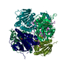
| Title | Molybdenum storage protein room-temperature structure determined by serial millisecond crystallography | ||||||
 Components Components | (Molybdenum storage protein subunit ...) x 2 | ||||||
 Keywords Keywords | HYDROLASE / room-temperature / serial crystallography | ||||||
| Function / homology |  Function and homology information Function and homology information | ||||||
| Biological species |  Azotobacter vinelandii DJ (bacteria) Azotobacter vinelandii DJ (bacteria) | ||||||
| Method |  X-RAY DIFFRACTION / SYNCHROTRON / MOLECULAR REPLACEMENT / Resolution: 1.7 Å X-RAY DIFFRACTION / SYNCHROTRON / MOLECULAR REPLACEMENT / Resolution: 1.7 Å | ||||||
 Authors Authors | Steffen, B. / Weinert, T. / Ermler, U. / Standfuss, J. | ||||||
 Citation Citation | Journal: Nat Commun / Year: 2017 Title: Serial millisecond crystallography for routine room-temperature structure determination at synchrotrons. Authors: Weinert, T. / Olieric, N. / Cheng, R. / Brunle, S. / James, D. / Ozerov, D. / Gashi, D. / Vera, L. / Marsh, M. / Jaeger, K. / Dworkowski, F. / Panepucci, E. / Basu, S. / Skopintsev, P. / ...Authors: Weinert, T. / Olieric, N. / Cheng, R. / Brunle, S. / James, D. / Ozerov, D. / Gashi, D. / Vera, L. / Marsh, M. / Jaeger, K. / Dworkowski, F. / Panepucci, E. / Basu, S. / Skopintsev, P. / Dore, A.S. / Geng, T. / Cooke, R.M. / Liang, M. / Prota, A.E. / Panneels, V. / Nogly, P. / Ermler, U. / Schertler, G. / Hennig, M. / Steinmetz, M.O. / Wang, M. / Standfuss, J. | ||||||
| History |
|
- Structure visualization
| Structure viewer | Molecule: MolmilJmol/JSmol |
|---|
- Downloads & links
-Download
| PDBx/mmCIF format | 5o5w.cif.gz | 119.9 KB | Display | PDBx/mmCIF format |
|---|---|---|---|---|
| PDB format | pdb5o5w.ent.gz | 89.6 KB | Display | PDB format |
| PDBx/mmJSON format | 5o5w.json.gz | Tree view | PDBx/mmJSON format | |
| Others |  Other downloads Other downloads |
-Validation report
| Arichive directory | https://data.pdbj.org/pub/pdb/validation_reports/o5/5o5wftp://data.pdbj.org/pub/pdb/validation_reports/o5/5o5w | HTTPS FTP |
|---|
-Related structure data
| Related structure data | 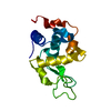 5njmC 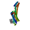 5nlxC 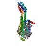 5nm2C 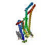 5nm4C  5nm5C  5nqtC  5nquC  4f6tS S: Starting model for refinement C: citing same article ( |
|---|---|
| Similar structure data |

-Links
 PDBj
PDBj- Assembly
| Deposited unit | 
| ||||||||||||
|---|---|---|---|---|---|---|---|---|---|---|---|---|---|
| 1 | 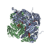
| ||||||||||||
| Unit cell |
| ||||||||||||
| Components on special symmetry positions |
|
-Components
-Molybdenum storage protein subunit ... , 2 types, 2 molecules AB
| #1: Protein | Mass: 29376.773 Da / Num. of mol.: 1 Source method: isolated from a genetically manipulated source Source: (gene. exp.) Azotobacter vinelandii DJ (bacteria) / Gene: mosA, Avin_43200 / Production host: |
|---|---|
| #2: Protein | Mass: 28378.775 Da / Num. of mol.: 1 Source method: isolated from a genetically manipulated source Source: (gene. exp.) Azotobacter vinelandii DJ (bacteria) / Gene: mosB, Avin_43210 / Production host: |
-Non-polymers , 6 types, 205 molecules 


| #3: Chemical | ChemComp-ATP /  Mass: 507.181 Da / Num. of mol.: 1 / Source method: obtained synthetically / Formula: C10H16N5O13P3 / Comment: ATP, energy-carrying molecule*YM Mass: 507.181 Da / Num. of mol.: 1 / Source method: obtained synthetically / Formula: C10H16N5O13P3 / Comment: ATP, energy-carrying molecule*YM | ||||||
|---|---|---|---|---|---|---|---|
| #4: Chemical | ChemComp-MG /  Mass: 24.305 Da / Num. of mol.: 1 / Source method: obtained synthetically / Formula: Mg Mass: 24.305 Da / Num. of mol.: 1 / Source method: obtained synthetically / Formula: Mg | ||||||
| #5: Chemical |  Mass: 1215.503 Da / Num. of mol.: 2 / Source method: obtained synthetically / Formula: Mo8O28 Mass: 1215.503 Da / Num. of mol.: 2 / Source method: obtained synthetically / Formula: Mo8O28#6: Chemical | ChemComp-M10 / ( |  Mass: 495.812 Da / Num. of mol.: 1 / Source method: obtained synthetically / Formula: Mo3O13 Mass: 495.812 Da / Num. of mol.: 1 / Source method: obtained synthetically / Formula: Mo3O13#7: Chemical | ChemComp-MO /  Mass: 95.940 Da / Num. of mol.: 22 / Source method: obtained synthetically / Formula: Mo Mass: 95.940 Da / Num. of mol.: 22 / Source method: obtained synthetically / Formula: Mo#8: Water | ChemComp-HOH / | Mass: 18.015 Da / Num. of mol.: 178 / Source method: isolated from a natural source / Formula: H2O |
-Experimental details
-Experiment
| Experiment | Method: X-RAY DIFFRACTION / Number of used crystals: 1 |
|---|
- Sample preparation
| Crystal grow | Temperature: 291 K / Method: vapor diffusion, hanging drop Details: 0,1 M Na-Citrat pH 5,6 1,4 M NH4H2PO4 20 % Glycerol |
|---|
-Data collection
| Diffraction | Mean temperature: 293 K |
|---|---|
| Diffraction source | Source: SYNCHROTRON / Site: SLS  / Beamline: X06SA / Wavelength: 1 Å / Beamline: X06SA / Wavelength: 1 Å |
| Detector | Type: DECTRIS EIGER X 16M / Detector: PIXEL / Date: Jun 1, 2016 |
| Radiation | Protocol: SINGLE WAVELENGTH / Monochromatic (M) / Laue (L): M / Scattering type: x-ray |
| Radiation wavelength | Wavelength: 1 Å / Relative weight: 1 |
| Reflection | Resolution: 1.7→24.2 Å / Num. obs: 110141 / % possible obs: 100 % / Redundancy: 765.4 % / Biso Wilson estimate: 29.42 Å2 / CC1/2: 0.99 / Rmerge(I) obs: 0.11 / Net I/σ(I): 5.29 |
- Processing
| Software |
| ||||||||||||||||||||||||||||||||||||||||||||||||||||||||||||||||||||||||||||||||||||||||||||||||||||||||||||||||||
|---|---|---|---|---|---|---|---|---|---|---|---|---|---|---|---|---|---|---|---|---|---|---|---|---|---|---|---|---|---|---|---|---|---|---|---|---|---|---|---|---|---|---|---|---|---|---|---|---|---|---|---|---|---|---|---|---|---|---|---|---|---|---|---|---|---|---|---|---|---|---|---|---|---|---|---|---|---|---|---|---|---|---|---|---|---|---|---|---|---|---|---|---|---|---|---|---|---|---|---|---|---|---|---|---|---|---|---|---|---|---|---|---|---|---|---|
| Refinement | Method to determine structure: MOLECULAR REPLACEMENT Starting model: 4F6T Resolution: 1.7→24.2 Å / Cor.coef. Fo:Fc: 0.964 / Cor.coef. Fo:Fc free: 0.958 / SU R Cruickshank DPI: 0.067 / Cross valid method: FREE R-VALUE / SU R Blow DPI: 0.067 / SU Rfree Blow DPI: 0.07 / SU Rfree Cruickshank DPI: 0.07
| ||||||||||||||||||||||||||||||||||||||||||||||||||||||||||||||||||||||||||||||||||||||||||||||||||||||||||||||||||
| Displacement parameters | Biso mean: 45 Å2
| ||||||||||||||||||||||||||||||||||||||||||||||||||||||||||||||||||||||||||||||||||||||||||||||||||||||||||||||||||
| Refine analyze | Luzzati coordinate error obs: 0.35 Å | ||||||||||||||||||||||||||||||||||||||||||||||||||||||||||||||||||||||||||||||||||||||||||||||||||||||||||||||||||
| Refinement step | Cycle: 1 / Resolution: 1.7→24.2 Å
| ||||||||||||||||||||||||||||||||||||||||||||||||||||||||||||||||||||||||||||||||||||||||||||||||||||||||||||||||||
| Refine LS restraints |
| ||||||||||||||||||||||||||||||||||||||||||||||||||||||||||||||||||||||||||||||||||||||||||||||||||||||||||||||||||
| LS refinement shell | Resolution: 1.7→1.74 Å / Rfactor Rfree error: 0 / Total num. of bins used: 20
|
