 Movie
Movie Controller

[English] 日本語
 Yorodumi
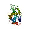
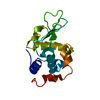
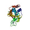
- PDB-1c10: CRYSTAL STRUCTURE OF HEW LYSOZYME UNDER PRESSURE OF XENON (8 BAR) -
+ Open data

- Basic information
| Entry | Database: PDB / ID: 1c10 | ||||||
|---|---|---|---|---|---|---|---|
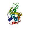
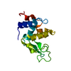
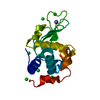
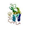
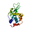
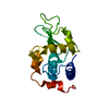
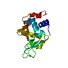
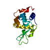
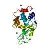
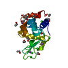
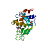
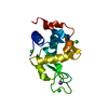
| Title | CRYSTAL STRUCTURE OF HEW LYSOZYME UNDER PRESSURE OF XENON (8 BAR) | ||||||
 Components Components | PROTEIN (LYSOZYME) | ||||||
 Keywords Keywords | HYDROLASE / HYDROPHOBIC CAVITY / XENON COMPLEX | ||||||
| Function / homology |  Function and homology information Function and homology informationLactose synthesis / Antimicrobial peptides / Neutrophil degranulation / beta-N-acetylglucosaminidase activity / cell wall macromolecule catabolic process / lysozyme / lysozyme activity / killing of cells of another organism / defense response to Gram-negative bacterium / defense response to bacterium ...Lactose synthesis / Antimicrobial peptides / Neutrophil degranulation / beta-N-acetylglucosaminidase activity / cell wall macromolecule catabolic process / lysozyme / lysozyme activity / killing of cells of another organism / defense response to Gram-negative bacterium / defense response to bacterium / defense response to Gram-positive bacterium / endoplasmic reticulum / : / identical protein binding / cytoplasm Similarity search - Function | ||||||
| Biological species |  | ||||||
| Method |  X-RAY DIFFRACTION / SYNCHROTRON / OTHER / Resolution: 2.03 Å X-RAY DIFFRACTION / SYNCHROTRON / OTHER / Resolution: 2.03 Å | ||||||
 Authors Authors | Prange, T. / Schiltz, M. / Pernot, L. / Colloc'h, N. / Longhi, S. / Bourguet, W. / Fourme, R. | ||||||
 Citation Citation | Journal: Proteins / Year: 1998 Title: Exploring hydrophobic sites in proteins with xenon or krypton. Authors: Prange, T. / Schiltz, M. / Pernot, L. / Colloc'h, N. / Longhi, S. / Bourguet, W. / Fourme, R. #1: Journal: Biochem.Biophys.Res.Commun. / Year: 1995Title: The Influence of Temperature on Lysozyme Crystals. Structure and Dynamics of Protein and Water Authors: Kurinov, I.V. / Harrison, R.W. | ||||||
| History |
|
- Structure visualization
| Structure viewer | Molecule: MolmilJmol/JSmol |
|---|
- Downloads & links
-Download
| PDBx/mmCIF format | 1c10.cif.gz | 40.3 KB | Display | PDBx/mmCIF format |
|---|---|---|---|---|
| PDB format | pdb1c10.ent.gz | 26.9 KB | Display | PDB format |
| PDBx/mmJSON format | 1c10.json.gz | Tree view | PDBx/mmJSON format | |
| Others |  Other downloads Other downloads |
-Validation report
| Arichive directory | https://data.pdbj.org/pub/pdb/validation_reports/c1/1c10ftp://data.pdbj.org/pub/pdb/validation_reports/c1/1c10 | HTTPS FTP |
|---|
-Related structure data
| Related structure data | 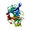 1c1mC  1c3lC 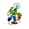 1qtkC 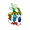 1lseS S: Starting model for refinement C: citing same article ( |
|---|---|
| Similar structure data |


-Links
 PDBj
PDBj


- Assembly
| Deposited unit | 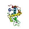
| ||||||||
|---|---|---|---|---|---|---|---|---|---|
| 1 |
| ||||||||
| Unit cell |
| ||||||||
| Components on special symmetry positions |
|
-Components
| #1: Protein | Mass: 14331.160 Da / Num. of mol.: 1 / Source method: isolated from a natural source / Source: (natural) | ||||||||
|---|---|---|---|---|---|---|---|---|---|
| #2: Chemical | ChemComp-NA /   Mass: 22.990 Da / Num. of mol.: 1 / Source method: obtained synthetically / Formula: Na Mass: 22.990 Da / Num. of mol.: 1 / Source method: obtained synthetically / Formula: Na | ||||||||
| #3: Chemical | ChemComp-CL /   Mass: 35.453 Da / Num. of mol.: 6 / Source method: obtained synthetically / Formula: Cl Mass: 35.453 Da / Num. of mol.: 6 / Source method: obtained synthetically / Formula: Cl#4: Chemical |   Mass: 131.293 Da / Num. of mol.: 3 / Source method: obtained synthetically / Formula: Xe Mass: 131.293 Da / Num. of mol.: 3 / Source method: obtained synthetically / Formula: Xe#5: Water | ChemComp-HOH / |  Mass: 18.015 Da / Num. of mol.: 69 / Source method: isolated from a natural source / Formula: H2O Mass: 18.015 Da / Num. of mol.: 69 / Source method: isolated from a natural source / Formula: H2OHas protein modification | Y | Nonpolymer details | OCCUPANCY FACTORS WERE REFINED. THE FIRST SITE IS ON A SPECIAL POSSITION AND HENCE REAL OCCUPANCY ...OCCUPANCY FACTORS WERE REFINED. THE FIRST SITE IS ON A SPECIAL POSSITION AND HENCE REAL OCCUPANCY IS TWICE THE REFINED VALUE | |
-Experimental details
-Experiment
| Experiment | Method: X-RAY DIFFRACTION / Number of used crystals: 1 |
|---|
- Sample preparation
| Crystal | Density Matthews: 2.03 Å3/Da / Density % sol: 39.4 % | ||||||||||||||||||||
|---|---|---|---|---|---|---|---|---|---|---|---|---|---|---|---|---|---|---|---|---|---|
| Crystal grow | pH: 5.5 / Details: SODIUM CHLORIDE, PH = 5.5 | ||||||||||||||||||||
| Crystal grow | *PLUS Temperature: 20 ℃ / pH: 5 / Method: unknown | ||||||||||||||||||||
| Components of the solutions | *PLUS
|
-Data collection
| Diffraction | Mean temperature: 277 K |
|---|---|
| Diffraction source | Source: SYNCHROTRON / Site: LURE  / Beamline: DW32 / Wavelength: 0.961 / Beamline: DW32 / Wavelength: 0.961 |
| Detector | Type: MARRESEARCH / Detector: IMAGE PLATE / Date: Feb 8, 1996 / Details: MULTILAYER MIRROR |
| Radiation | Monochromator: SI (III) / Protocol: SINGLE WAVELENGTH / Monochromatic (M) / Laue (L): M / Scattering type: x-ray |
| Radiation wavelength | Wavelength: 0.961 Å / Relative weight: 1 |
| Reflection | Resolution: 2.03→9.9 Å / Num. obs: 6454 / % possible obs: 79 % / Observed criterion σ(I): 4 / Redundancy: 5.1 % / Rmerge(I) obs: 0.044 / Net I/σ(I): 8.9 |
| Reflection shell | Resolution: 2.03→2.15 Å / Redundancy: 2.9 % / Rmerge(I) obs: 0.152 / Mean I/σ(I) obs: 4.3 / % possible all: 59 |
| Reflection shell | *PLUS % possible obs: 59 % |
- Processing
| Software |
| |||||||||||||||||||||||||||||||||
|---|---|---|---|---|---|---|---|---|---|---|---|---|---|---|---|---|---|---|---|---|---|---|---|---|---|---|---|---|---|---|---|---|---|---|
| Refinement | Method to determine structure: OTHER Starting model: PDB ENTRY 1LSE Resolution: 2.03→9.9 Å / Num. parameters: 4285 / Num. restraintsaints: 4086 / σ(F): 4 / Stereochemistry target values: ENGH & HUBER / Details: USED WEIGHTED FULL MATRIX LEAST SQUARES PROCEDURE
| |||||||||||||||||||||||||||||||||
| Refine analyze | Num. disordered residues: 0 | |||||||||||||||||||||||||||||||||
| Refinement step | Cycle: LAST / Resolution: 2.03→9.9 Å
| |||||||||||||||||||||||||||||||||
| Refine LS restraints |
|
