 Movie
Movie Controller


+ Open data

- Basic information
| Entry | Database: PDB / ID: 1c3l | ||||||
|---|---|---|---|---|---|---|---|
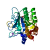
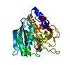
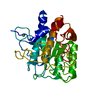
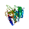
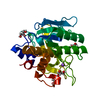
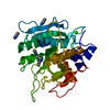
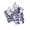
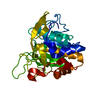
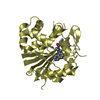
| Title | SUBTILISIN-CARLSBERG COMPLEXED WITH XENON (8 BAR) | ||||||
 Components Components | SUBTILISIN-CARLSBERG | ||||||
 Keywords Keywords | HYDROLASE / XENON / SERINE-PROTEINASE | ||||||
| Function / homology |  Function and homology information Function and homology informationsubtilisin / serine-type endopeptidase activity / proteolysis / : / metal ion binding Similarity search - Function | ||||||
| Biological species |  | ||||||
| Method |  X-RAY DIFFRACTION / SYNCHROTRON / Resolution: 2.16 Å X-RAY DIFFRACTION / SYNCHROTRON / Resolution: 2.16 Å | ||||||
 Authors Authors | Prange, T. / Schiltz, M. / Pernot, L. / Colloc'h, N. / Longhi, S. | ||||||
 Citation Citation | Journal: Proteins / Year: 1998 Title: Exploring hydrophobic sites in proteins with xenon or krypton. Authors: Prange, T. / Schiltz, M. / Pernot, L. / Colloc'h, N. / Longhi, S. / Bourguet, W. / Fourme, R. #1: Journal: J.Appl.Crystallogr. / Year: 1994Title: On the Preparation and X-ray Data Collection of Isomorphous Xenon Derivatives Authors: Schiltz, M. / Prange, T. / Fourme, R. #2: Journal: Structure / Year: 1995Title: The Catalytic Site of Serine-Proteinases as a Specific Binding Cavity for Xenon Authors: Schiltz, M. / Fourme, R. / Broutin, I. / Prange, T. #3: Journal: J.Mol.Biol. / Year: 1976Title: The Structure of Subtilopeptidase 1. X-ray Crystallographic Data Authors: Petsko, G.A. / Tsernoglou, D. | ||||||
| History |
|
- Structure visualization
| Structure viewer | Molecule: MolmilJmol/JSmol |
|---|
- Downloads & links
-Download
| PDBx/mmCIF format | 1c3l.cif.gz | 64.6 KB | Display | PDBx/mmCIF format |
|---|---|---|---|---|
| PDB format | pdb1c3l.ent.gz | 45.9 KB | Display | PDB format |
| PDBx/mmJSON format | 1c3l.json.gz | Tree view | PDBx/mmJSON format | |
| Others |  Other downloads Other downloads |
-Validation report
| Arichive directory | https://data.pdbj.org/pub/pdb/validation_reports/c3/1c3lftp://data.pdbj.org/pub/pdb/validation_reports/c3/1c3l | HTTPS FTP |
|---|
-Related structure data
| Related structure data | 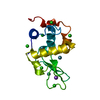 1c10C 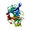 1c1mC 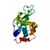 1qtkC 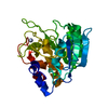 1scaS S: Starting model for refinement C: citing same article ( |
|---|---|
| Similar structure data |


-Links
 PDBj
PDBj


- Assembly
| Deposited unit | 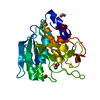
| ||||||||||
|---|---|---|---|---|---|---|---|---|---|---|---|
| 1 |
| ||||||||||
| Unit cell |
|
-Components
| #1: Protein | Mass: 27279.176 Da / Num. of mol.: 1 / Source method: isolated from a natural source / Source: (natural) | ||||||
|---|---|---|---|---|---|---|---|
| #2: Chemical |   Mass: 40.078 Da / Num. of mol.: 2 / Source method: obtained synthetically / Formula: Ca Mass: 40.078 Da / Num. of mol.: 2 / Source method: obtained synthetically / Formula: Ca#3: Chemical | ChemComp-XE / |   Mass: 131.293 Da / Num. of mol.: 1 / Source method: obtained synthetically / Formula: Xe Mass: 131.293 Da / Num. of mol.: 1 / Source method: obtained synthetically / Formula: Xe#4: Chemical |   Mass: 46.025 Da / Num. of mol.: 3 / Source method: obtained synthetically / Formula: CH2O2 Mass: 46.025 Da / Num. of mol.: 3 / Source method: obtained synthetically / Formula: CH2O2#5: Water | ChemComp-HOH / |  Mass: 18.015 Da / Num. of mol.: 108 / Source method: isolated from a natural source / Formula: H2O Mass: 18.015 Da / Num. of mol.: 108 / Source method: isolated from a natural source / Formula: H2O |
-Experimental details
-Experiment
| Experiment | Method: X-RAY DIFFRACTION / Number of used crystals: 1 |
|---|
- Sample preparation
| Crystal | Density Matthews: 2.07 Å3/Da / Density % sol: 40.57 % | ||||||||||||||||||||
|---|---|---|---|---|---|---|---|---|---|---|---|---|---|---|---|---|---|---|---|---|---|
| Crystal grow | Temperature: 298 K / Method: microdialysis / pH: 5.5 Details: protein: 15mg/ml in acetate buffer (0.05M) plus sodium azide (10-5M). Sodium formate as the crystallizing agent(4M). Crystals as elongated needles in two weeks., pH 5.50, MICRODIALYSIS, temperature 298K | ||||||||||||||||||||
| Crystal grow | *PLUS Temperature: 20 ℃ / pH: 5 / Method: unknown / Details: Shotton, D.M., (1968) J.Mol.Biol., 32, 155. | ||||||||||||||||||||
| Components of the solutions | *PLUS
|
-Data collection
| Diffraction | Mean temperature: 278 K |
|---|---|
| Diffraction source | Source: SYNCHROTRON / Site: LURE  / Beamline: DW32 / Wavelength: 0.961 / Beamline: DW32 / Wavelength: 0.961 |
| Detector | Type: MARRESEARCH / Detector: IMAGE PLATE / Date: Oct 20, 1995 |
| Radiation | Protocol: SINGLE WAVELENGTH / Monochromatic (M) / Laue (L): M / Scattering type: x-ray |
| Radiation wavelength | Wavelength: 0.961 Å / Relative weight: 1 |
| Reflection | Resolution: 2.16→15 Å / Num. all: 44356 / Num. obs: 40233 / % possible obs: 0.83 % / Observed criterion σ(F): 3 / Observed criterion σ(I): 4 / Redundancy: 4.1 % / Biso Wilson estimate: 24 Å2 / Rmerge(I) obs: 0.062 / Net I/σ(I): 11 |
| Reflection shell | Resolution: 2.16→2.27 Å / Redundancy: 2.8 % / Rmerge(I) obs: 0.167 / Num. unique all: 1172 / % possible all: 65 |
- Processing
| Software |
| |||||||||||||||||||||||||
|---|---|---|---|---|---|---|---|---|---|---|---|---|---|---|---|---|---|---|---|---|---|---|---|---|---|---|
| Refinement | Starting model: 1SCA Resolution: 2.16→7 Å / Num. parameters: 8135 / Num. restraintsaints: 7863 / σ(F): 3 / σ(I): 4 / Stereochemistry target values: Engh & Huber / Details: 8135 parameters refined using 7863 restraints
| |||||||||||||||||||||||||
| Refinement step | Cycle: LAST / Resolution: 2.16→7 Å
| |||||||||||||||||||||||||
| Refine LS restraints |
| |||||||||||||||||||||||||
| Software | *PLUS Name: SHELXL-97 / Classification: refinement | |||||||||||||||||||||||||
| Refinement | *PLUS σ(F): 3 | |||||||||||||||||||||||||
| Solvent computation | *PLUS | |||||||||||||||||||||||||
| Displacement parameters | *PLUS |
