 Movie
Movie Controller


+ Open data

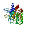
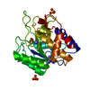
- Basic information
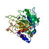
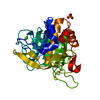
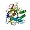
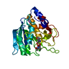
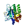
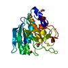
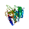
| Entry | Database: PDB / ID: 1bfk | ||||||
|---|---|---|---|---|---|---|---|
| Title | CRYSTAL STRUCTURE OF SUBTILISIN CARLSBERG IN 40% ACETONITRILE | ||||||
 Components Components | SUBTILISIN CARLSBERG | ||||||
 Keywords Keywords | SERINE PROTEASE / ORGANIC SOLVENT / MIXTURES | ||||||
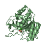
| Function / homology |  Function and homology information Function and homology informationsubtilisin / serine-type endopeptidase activity / proteolysis / : / metal ion binding Similarity search - Function | ||||||
| Biological species |  | ||||||
| Method |  X-RAY DIFFRACTION / MOLECULAR REPLACEMENT / Resolution: 2.3 Å X-RAY DIFFRACTION / MOLECULAR REPLACEMENT / Resolution: 2.3 Å | ||||||
 Authors Authors | Schmitke, J.L. / Stern, L.J. / Klibanov, A.M. | ||||||
 Citation Citation | Journal: Biochem.Biophys.Res.Commun. / Year: 1998 Title: Organic solvent binding to crystalline subtilisin1 in mostly aqueous media and in the neat solvents. Authors: Schmitke, J.L. / Stern, L.J. / Klibanov, A.M. | ||||||
| History |
|
- Structure visualization
| Structure viewer | Molecule: MolmilJmol/JSmol |
|---|
- Downloads & links
-Download
| PDBx/mmCIF format | 1bfk.cif.gz | 62.8 KB | Display | PDBx/mmCIF format |
|---|---|---|---|---|
| PDB format | pdb1bfk.ent.gz | 45 KB | Display | PDB format |
| PDBx/mmJSON format | 1bfk.json.gz | Tree view | PDBx/mmJSON format | |
| Others |  Other downloads Other downloads |
-Validation report
| Arichive directory | https://data.pdbj.org/pub/pdb/validation_reports/bf/1bfkftp://data.pdbj.org/pub/pdb/validation_reports/bf/1bfk | HTTPS FTP |
|---|
-Related structure data
| Related structure data |  1bfuC 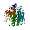 1scbS S: Starting model for refinement C: citing same article ( |
|---|---|
| Similar structure data |


-Links
 PDBj
PDBj

- Assembly
| Deposited unit | 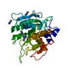
| ||||||||
|---|---|---|---|---|---|---|---|---|---|
| 1 |
| ||||||||
| Unit cell |
|
-Components
| #1: Protein | Mass: 27306.199 Da / Num. of mol.: 1 / Source method: isolated from a natural source / Source: (natural) | ||
|---|---|---|---|
| #2: Chemical | ChemComp-CA /   Mass: 40.078 Da / Num. of mol.: 1 / Source method: obtained synthetically / Formula: Ca Mass: 40.078 Da / Num. of mol.: 1 / Source method: obtained synthetically / Formula: Ca | ||
| #3: Chemical | ChemComp-CCN /   Mass: 41.052 Da / Num. of mol.: 5 / Source method: obtained synthetically / Formula: C2H3N Mass: 41.052 Da / Num. of mol.: 5 / Source method: obtained synthetically / Formula: C2H3N#4: Water | ChemComp-HOH / |  Mass: 18.015 Da / Num. of mol.: 74 / Source method: isolated from a natural source / Formula: H2O Mass: 18.015 Da / Num. of mol.: 74 / Source method: isolated from a natural source / Formula: H2O |
-Experimental details
-Experiment
| Experiment | Method: X-RAY DIFFRACTION / Number of used crystals: 2 |
|---|
- Sample preparation
| Crystal | Density Matthews: 2.1 Å3/Da / Density % sol: 42 % | |||||||||||||||
|---|---|---|---|---|---|---|---|---|---|---|---|---|---|---|---|---|
| Crystal grow | pH: 5.6 Details: PROTEIN WAS CRYSTALLIZED FROM 13% SODIUM SULFATE, 30 MM CACODYLATE, PH 5.6; THEN CROSS-LINKED WITH 10% GLUTARALDEHYDE IN 13% SODIUM SULFATE, 30 MM CACODYLATE, PH 7.5, WASHED WITH DISTILLED ...Details: PROTEIN WAS CRYSTALLIZED FROM 13% SODIUM SULFATE, 30 MM CACODYLATE, PH 5.6; THEN CROSS-LINKED WITH 10% GLUTARALDEHYDE IN 13% SODIUM SULFATE, 30 MM CACODYLATE, PH 7.5, WASHED WITH DISTILLED WATER, AND TRANSFERRED TO AN AQUEOUS SYSTEM CONTAINING 40% ACETONITRILE. | |||||||||||||||
| Crystal grow | *PLUS Method: unknown | |||||||||||||||
| Components of the solutions | *PLUS
|
-Data collection
| Diffraction | Mean temperature: 296 K |
|---|---|
| Diffraction source | Source: ROTATING ANODE / Type: RIGAKU RUH2R / Wavelength: 1.5418 |
| Detector | Type: RIGAKU RAXIS II / Detector: IMAGE PLATE / Date: Nov 19, 1997 / Details: MIRROR |
| Radiation | Monochromatic (M) / Laue (L): M / Scattering type: x-ray |
| Radiation wavelength | Wavelength: 1.5418 Å / Relative weight: 1 |
| Reflection | Resolution: 2.3→20 Å / Num. obs: 10215 / % possible obs: 93.7 % / Redundancy: 3.3 % / Biso Wilson estimate: 35 Å2 / Rsym value: 0.126 / Net I/σ(I): 10 |
| Reflection shell | Resolution: 2.3→2.4 Å / Redundancy: 3.2 % / Mean I/σ(I) obs: 3.9 / Rsym value: 0.406 / % possible all: 85.9 |
| Reflection | *PLUS Lowest resolution: 20 Å / Num. measured all: 34019 / Rmerge(I) obs: 0.126 |
| Reflection shell | *PLUS % possible obs: 85.9 % / Num. unique obs: 1098 / Num. measured obs: 3523 / Rmerge(I) obs: 0.406 |
- Processing
| Software |
| ||||||||||||||||||||||||||||||||||||||||||||||||||||||||||||
|---|---|---|---|---|---|---|---|---|---|---|---|---|---|---|---|---|---|---|---|---|---|---|---|---|---|---|---|---|---|---|---|---|---|---|---|---|---|---|---|---|---|---|---|---|---|---|---|---|---|---|---|---|---|---|---|---|---|---|---|---|---|
| Refinement | Method to determine structure: MOLECULAR REPLACEMENT Starting model: 1SCB Resolution: 2.3→20 Å / Data cutoff high absF: 100000 / Data cutoff low absF: 0.1 / Isotropic thermal model: RESTRAINED / σ(F): 2
| ||||||||||||||||||||||||||||||||||||||||||||||||||||||||||||
| Displacement parameters | Biso mean: 28 Å2 | ||||||||||||||||||||||||||||||||||||||||||||||||||||||||||||
| Refine analyze | Luzzati coordinate error obs: 0.31 Å / Luzzati d res low obs: 5 Å / Luzzati sigma a obs: 0.35 Å | ||||||||||||||||||||||||||||||||||||||||||||||||||||||||||||
| Refinement step | Cycle: LAST / Resolution: 2.3→20 Å
| ||||||||||||||||||||||||||||||||||||||||||||||||||||||||||||
| Refine LS restraints |
| ||||||||||||||||||||||||||||||||||||||||||||||||||||||||||||
| LS refinement shell | Resolution: 2.3→2.4 Å / Total num. of bins used: 8
| ||||||||||||||||||||||||||||||||||||||||||||||||||||||||||||
| Xplor file |
| ||||||||||||||||||||||||||||||||||||||||||||||||||||||||||||
| Software | *PLUS Name: X-PLOR / Version: 3.8 / Classification: refinement | ||||||||||||||||||||||||||||||||||||||||||||||||||||||||||||
| Refinement | *PLUS σ(F): 2 | ||||||||||||||||||||||||||||||||||||||||||||||||||||||||||||
| Solvent computation | *PLUS | ||||||||||||||||||||||||||||||||||||||||||||||||||||||||||||
| Displacement parameters | *PLUS | ||||||||||||||||||||||||||||||||||||||||||||||||||||||||||||
| Refine LS restraints | *PLUS
| ||||||||||||||||||||||||||||||||||||||||||||||||||||||||||||
| LS refinement shell | *PLUS Rfactor obs: 0.284 |
