 Movie
Movie Controller

[English] 日本語
 Yorodumi
- PDB-3qtl: Structural Basis for Dual-inhibition Mechanism of a Non-classical... -
+ Open data

- Basic information
| Entry | Database: PDB / ID: 3qtl | ||||||
|---|---|---|---|---|---|---|---|
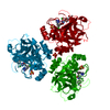
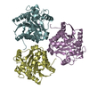
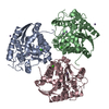
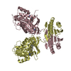
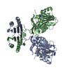
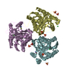
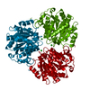
| Title | Structural Basis for Dual-inhibition Mechanism of a Non-classical Kazal-type Serine Protease Inhibitor from Horseshoe Crab in Complex with Subtilisin | ||||||
 Components Components |
| ||||||
 Keywords Keywords | HYDROLASE/HYDROLASE INHIBITOR / SERINE PROTEASE -KAZAL TYPE SERINE PROTEASE INHIBITOR COMPLEX / HYDROLASE INHIBITOR / HYDROLASE-HYDROLASE INHIBITOR complex | ||||||
| Function / homology |  Function and homology information Function and homology informationserine-type endopeptidase activity / proteolysis / : / metal ion binding Similarity search - Function | ||||||
| Biological species |   Carcinoscorpius rotundicauda (Southeast Asian horseshoe crab) Carcinoscorpius rotundicauda (Southeast Asian horseshoe crab) | ||||||
| Method |  X-RAY DIFFRACTION / SYNCHROTRON / MOLECULAR REPLACEMENT / molecular replacement / Resolution: 2.6 Å X-RAY DIFFRACTION / SYNCHROTRON / MOLECULAR REPLACEMENT / molecular replacement / Resolution: 2.6 Å | ||||||
 Authors Authors | Shenoy, R.T. / Sivaraman, J. | ||||||
 Citation Citation | Journal: Plos One / Year: 2011 Title: Structural basis for dual-inhibition mechanism of a non-classical kazal-type serine protease inhibitor from horseshoe crab in complex with subtilisin. Authors: Shenoy, R.T. / Thangamani, S. / Velazquez-Campoy, A. / Ho, B. / Ding, J.L. / Sivaraman, J. | ||||||
| History |
|
- Structure visualization
| Structure viewer | Molecule: MolmilJmol/JSmol |
|---|
- Downloads & links
-Download
| PDBx/mmCIF format | 3qtl.cif.gz | 166.1 KB | Display | PDBx/mmCIF format |
|---|---|---|---|---|
| PDB format | pdb3qtl.ent.gz | 131.7 KB | Display | PDB format |
| PDBx/mmJSON format | 3qtl.json.gz | Tree view | PDBx/mmJSON format | |
| Others |  Other downloads Other downloads |
-Validation report
| Arichive directory | https://data.pdbj.org/pub/pdb/validation_reports/qt/3qtlftp://data.pdbj.org/pub/pdb/validation_reports/qt/3qtl | HTTPS FTP |
|---|
-Related structure data
| Related structure data | 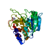 1scaS S: Starting model for refinement |
|---|---|
| Similar structure data |

-Links
 PDBj
PDBj


- Assembly
| Deposited unit | 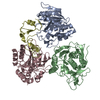
| ||||||||
|---|---|---|---|---|---|---|---|---|---|
| 1 |
| ||||||||
| 2 | 
| ||||||||
| 3 | 
| ||||||||
| Unit cell |
|
-Components
| #1: Protein | Mass: 27306.199 Da / Num. of mol.: 3 / Fragment: UNP RESIDUES 106-379 Source method: isolated from a genetically manipulated source Source: (gene. exp.) References: UniProt: Q1EM64, UniProt: Q6PNN5*PLUS, subtilisin #2: Protein | | Mass: 8311.360 Da / Num. of mol.: 1 / Fragment: UNP RESIDUES 24-99 Source method: isolated from a genetically manipulated source Source: (gene. exp.) Carcinoscorpius rotundicauda (Southeast Asian horseshoe crab)Gene: SPI-1 / Plasmid: pET32b(+) / Production host: #3: Water | ChemComp-HOH / |  Mass: 18.015 Da / Num. of mol.: 157 / Source method: isolated from a natural source / Formula: H2O Mass: 18.015 Da / Num. of mol.: 157 / Source method: isolated from a natural source / Formula: H2OHas protein modification | Y | |
|---|
-Experimental details
-Experiment
| Experiment | Method: X-RAY DIFFRACTION / Number of used crystals: 1 |
|---|
- Sample preparation
| Crystal | Density Matthews: 2.97 Å3/Da / Density % sol: 58.53 % |
|---|---|
| Crystal grow | Temperature: 298 K / Method: vapor diffusion, hanging drop / pH: 6.5 Details: 11% (w/v) PEG 20000, 0.1M 2-(N-morpholino)ethanesulfonic acid (MES) pH 6.5, VAPOR DIFFUSION, HANGING DROP, temperature 298.0K |
-Data collection
| Diffraction | Mean temperature: 298 K |
|---|---|
| Diffraction source | Source: SYNCHROTRON / Site: NSLS  / Beamline: X29A / Wavelength: 1.5418 Å / Beamline: X29A / Wavelength: 1.5418 Å |
| Detector | Date: Sep 29, 2007 |
| Radiation | Monochromator: Rosenbaum-Rock double crystal sagittal focusing monochromator Protocol: SINGLE WAVELENGTH / Monochromatic (M) / Laue (L): M / Scattering type: x-ray |
| Radiation wavelength | Wavelength: 1.5418 Å / Relative weight: 1 |
| Reflection | Resolution: 2.6→15 Å / Num. obs: 32884 / % possible obs: 99.8 % / Observed criterion σ(F): 2 / Observed criterion σ(I): 2 / Redundancy: 6.3 % / Rsym value: 0.1 |
| Reflection shell | Resolution: 2.6→2.64 Å / Redundancy: 5.3 % / Rsym value: 0.34 / % possible all: 99.3 |
-Phasing
| Phasing | Method: molecular replacement |
|---|
- Processing
| Software |
| ||||||||||||||||||||||||||||||||||||||||||||||||||||||||||||||||||
|---|---|---|---|---|---|---|---|---|---|---|---|---|---|---|---|---|---|---|---|---|---|---|---|---|---|---|---|---|---|---|---|---|---|---|---|---|---|---|---|---|---|---|---|---|---|---|---|---|---|---|---|---|---|---|---|---|---|---|---|---|---|---|---|---|---|---|---|
| Refinement | Method to determine structure: MOLECULAR REPLACEMENT Starting model: PDB ENTRY 1SCA Resolution: 2.6→14.901 Å / SU ML: 0.36 / σ(F): 0.14 / Phase error: 24.46 / Stereochemistry target values: ML
| ||||||||||||||||||||||||||||||||||||||||||||||||||||||||||||||||||
| Solvent computation | Shrinkage radii: 0.9 Å / VDW probe radii: 1.11 Å / Bsol: 44.037 Å2 / ksol: 0.431 e/Å3 | ||||||||||||||||||||||||||||||||||||||||||||||||||||||||||||||||||
| Displacement parameters |
| ||||||||||||||||||||||||||||||||||||||||||||||||||||||||||||||||||
| Refinement step | Cycle: LAST / Resolution: 2.6→14.901 Å
| ||||||||||||||||||||||||||||||||||||||||||||||||||||||||||||||||||
| Refine LS restraints |
| ||||||||||||||||||||||||||||||||||||||||||||||||||||||||||||||||||
| LS refinement shell | Refine-ID: X-RAY DIFFRACTION / Total num. of bins used: 10
|
