 Movie
Movie Controller


+ Open data

- Basic information
| Entry | Database: PDB / ID: 1xs0 | ||||||
|---|---|---|---|---|---|---|---|
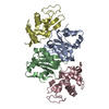
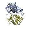
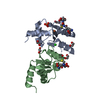
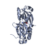
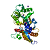
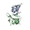
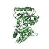
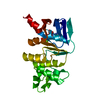
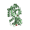
| Title | Structure of the E. coli Ivy protein | ||||||
 Components Components | Inhibitor of vertebrate lysozyme | ||||||
 Keywords Keywords | HYDROLASE INHIBITOR / alpha beta fold / dimer | ||||||
| Function / homology |  Function and homology information Function and homology informationlysozyme inhibitor activity / negative regulation of catalytic activity / outer membrane-bounded periplasmic space / periplasmic space / protein homodimerization activity Similarity search - Function | ||||||
| Biological species |  | ||||||
| Method |  X-RAY DIFFRACTION / SYNCHROTRON / MAD / Resolution: 1.58 Å X-RAY DIFFRACTION / SYNCHROTRON / MAD / Resolution: 1.58 Å | ||||||
 Authors Authors | Abergel, C. / Monchois, V. / Byrn, D. / Lazzaroni, J.C. / Claverie, J.M. | ||||||
 Citation Citation | Journal: Proc.Natl.Acad.Sci.USA / Year: 2007 Title: Structure and evolution of the Ivy protein family, unexpected lysozyme inhibitors in Gram-negative bacteria. Authors: Abergel, C. / Monchois, V. / Byrne, D. / Chenivesse, S. / Lembo, F. / Lazzaroni, J.C. / Claverie, J.M. #1: Journal: J.Biol.Chem. / Year: 2001 Title: Escherichia coli ykfE ORFan gene encodes a potent inhibitor of C-type lysozyme Authors: Monchois, V. / Abergel, C. / Sturgis, J. / Jeudy, S. / Claverie, J.M. #2: Journal: ACTA CRYSTALLOGR.,SECT.D / Year: 2000 Title: Crystallization and preliminary crystallographic study of b0220, an 'ORFan' protein of unknown function from Escherichia coli Authors: Abergel, C. / Monchois, V. / Chenivesse, S. / Jeudy, S. / Claverie, J.M. | ||||||
| History |
|
- Structure visualization
| Structure viewer | Molecule: MolmilJmol/JSmol |
|---|
- Downloads & links
-Download
| PDBx/mmCIF format | 1xs0.cif.gz | 90.9 KB | Display | PDBx/mmCIF format |
|---|---|---|---|---|
| PDB format | pdb1xs0.ent.gz | 69.7 KB | Display | PDB format |
| PDBx/mmJSON format | 1xs0.json.gz | Tree view | PDBx/mmJSON format | |
| Others |  Other downloads Other downloads |
-Validation report
| Arichive directory | https://data.pdbj.org/pub/pdb/validation_reports/xs/1xs0ftp://data.pdbj.org/pub/pdb/validation_reports/xs/1xs0 | HTTPS FTP |
|---|
-Related structure data


-Links
 PDBj
PDBj- Assembly
| Deposited unit | 
| ||||||||
|---|---|---|---|---|---|---|---|---|---|
| 1 | 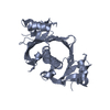
| ||||||||
| 2 | 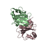
| ||||||||
| Unit cell |
| ||||||||
| Components on special symmetry positions |
|
-Components
| #1: Protein | Mass: 15060.833 Da / Num. of mol.: 3 Source method: isolated from a genetically manipulated source Source: (gene. exp.) #2: Water | ChemComp-HOH / |  Mass: 18.015 Da / Num. of mol.: 302 / Source method: isolated from a natural source / Formula: H2O Mass: 18.015 Da / Num. of mol.: 302 / Source method: isolated from a natural source / Formula: H2OHas protein modification | Y | |
|---|
-Experimental details
-Experiment
| Experiment | Method: X-RAY DIFFRACTION / Number of used crystals: 1 |
|---|
- Sample preparation
| Crystal | Density Matthews: 1.99 Å3/Da / Density % sol: 38 % |
|---|---|
| Crystal grow | Temperature: 293 K / Method: vapor diffusion, hanging drop / pH: 6.75 Details: 1.4M sodium citrate, 8% PEG 8000, 20mM TRIS, 50mM NaCl, pH 6.75, VAPOR DIFFUSION, HANGING DROP, temperature 293K |
-Data collection
| Diffraction | Mean temperature: 105 K | ||||||||||||
|---|---|---|---|---|---|---|---|---|---|---|---|---|---|
| Diffraction source | Source: SYNCHROTRON / Site: ESRF  / Beamline: BM30A / Wavelength: 0.97917, 0.97941, 0.97393 / Beamline: BM30A / Wavelength: 0.97917, 0.97941, 0.97393 | ||||||||||||
| Detector | Type: MARRESEARCH / Detector: IMAGE PLATE / Date: May 30, 2000 | ||||||||||||
| Radiation | Protocol: MAD / Monochromatic (M) / Laue (L): M / Scattering type: x-ray | ||||||||||||
| Radiation wavelength |
| ||||||||||||
| Reflection | Resolution: 1.58→40.7 Å / Num. all: 44577 / Num. obs: 44577 / % possible obs: 98.1 % / Observed criterion σ(F): 1 / Observed criterion σ(I): 9.3 / Redundancy: 3 % / Biso Wilson estimate: 25.7 Å2 / Rsym value: 0.037 | ||||||||||||
| Reflection shell | Resolution: 1.58→1.67 Å / Redundancy: 2.5 % / Mean I/σ(I) obs: 3.1 / Num. unique all: 3539 / Rsym value: 0.223 / % possible all: 98.1 |
- Processing
| Software |
| |||||||||||||||||||||||||
|---|---|---|---|---|---|---|---|---|---|---|---|---|---|---|---|---|---|---|---|---|---|---|---|---|---|---|
| Refinement | Method to determine structure: MAD / Resolution: 1.58→14.52 Å / Rfactor Rfree error: 0.004 / Data cutoff high absF: 823714.1 / Data cutoff low absF: 0 / Isotropic thermal model: RESTRAINED / Cross valid method: THROUGHOUT / σ(F): 0 / Stereochemistry target values: Engh & Huber
| |||||||||||||||||||||||||
| Solvent computation | Solvent model: FLAT MODEL / Bsol: 60.5474 Å2 / ksol: 0.390667 e/Å3 | |||||||||||||||||||||||||
| Displacement parameters | Biso mean: 29.7 Å2
| |||||||||||||||||||||||||
| Refine analyze |
| |||||||||||||||||||||||||
| Refinement step | Cycle: LAST / Resolution: 1.58→14.52 Å
| |||||||||||||||||||||||||
| Refine LS restraints |
| |||||||||||||||||||||||||
| LS refinement shell | Resolution: 1.58→1.68 Å / Rfactor Rfree error: 0.011 / Total num. of bins used: 6
| |||||||||||||||||||||||||
| Xplor file |
|
