 Movie
Movie Controller

[English] 日本語
 Yorodumi
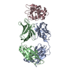
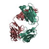
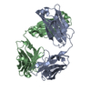
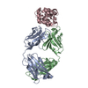
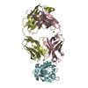
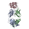
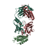
- PDB-1ndg: Crystal structure of Fab fragment of antibody HyHEL-8 complexed w... -
+ Open data

- Basic information
| Entry | Database: PDB / ID: 1ndg | ||||||
|---|---|---|---|---|---|---|---|
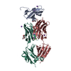
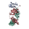
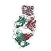
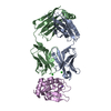
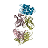
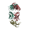
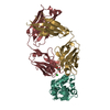
| Title | Crystal structure of Fab fragment of antibody HyHEL-8 complexed with its antigen lysozyme | ||||||
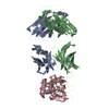
 Components Components |
| ||||||
 Keywords Keywords | IMMUNE SYSTEM/HYDROLASE / antibody / lysozyme / mutant / HyHEL-8 / IMMUNE SYSTEM-HYDROLASE COMPLEX | ||||||
| Function / homology |  Function and homology information Function and homology informationpositive regulation of type IIa hypersensitivity / positive regulation of B cell activation / humoral immune response mediated by circulating immunoglobulin / phagocytosis, recognition / early endosome to late endosome transport / positive regulation of type I hypersensitivity / antibody-dependent cellular cytotoxicity / Fc-gamma receptor I complex binding / alpha-beta T cell receptor complex / immunoglobulin complex, circulating ...positive regulation of type IIa hypersensitivity / positive regulation of B cell activation / humoral immune response mediated by circulating immunoglobulin / phagocytosis, recognition / early endosome to late endosome transport / positive regulation of type I hypersensitivity / antibody-dependent cellular cytotoxicity / Fc-gamma receptor I complex binding / alpha-beta T cell receptor complex / immunoglobulin complex, circulating / phagocytosis, engulfment / immunoglobulin receptor binding / antigen processing and presentation / IgG immunoglobulin complex / endosome to lysosome transport / immunoglobulin mediated immune response / regulation of proteolysis / complement activation, classical pathway / positive regulation of endocytosis / antigen binding / multivesicular body / B cell differentiation / Lactose synthesis / Antimicrobial peptides / positive regulation of phagocytosis / response to bacterium / Neutrophil degranulation / beta-N-acetylglucosaminidase activity / positive regulation of immune response / cell wall macromolecule catabolic process / lysozyme / lysozyme activity / antibacterial humoral response / killing of cells of another organism / defense response to Gram-negative bacterium / adaptive immune response / defense response to bacterium / defense response to Gram-positive bacterium / endoplasmic reticulum / : / extracellular region / identical protein binding / plasma membrane / cytoplasm Similarity search - Function | ||||||
| Biological species |   | ||||||
| Method |  X-RAY DIFFRACTION / SYNCHROTRON / MOLECULAR REPLACEMENT / Resolution: 1.9 Å X-RAY DIFFRACTION / SYNCHROTRON / MOLECULAR REPLACEMENT / Resolution: 1.9 Å | ||||||
 Authors Authors | Mariuzza, R.A. / Li, Y. / Li, H. / Yang, F. / Smith-Gill, S.J. | ||||||
 Citation Citation | Journal: Nat.Struct.Biol. / Year: 2003 Title: X-ray snapshots of the maturation of an antibody response to a protein antigen Authors: Li, Y. / Li, H. / Yang, F. / Smith-Gill, S.J. / Mariuzza, R.A. | ||||||
| History |
|
- Structure visualization
| Structure viewer | Molecule: MolmilJmol/JSmol |
|---|
- Downloads & links
-Download
| PDBx/mmCIF format | 1ndg.cif.gz | 129.6 KB | Display | PDBx/mmCIF format |
|---|---|---|---|---|
| PDB format | pdb1ndg.ent.gz | 100.3 KB | Display | PDB format |
| PDBx/mmJSON format | 1ndg.json.gz | Tree view | PDBx/mmJSON format | |
| Others |  Other downloads Other downloads |
-Validation report
| Arichive directory | https://data.pdbj.org/pub/pdb/validation_reports/nd/1ndgftp://data.pdbj.org/pub/pdb/validation_reports/nd/1ndg | HTTPS FTP |
|---|
-Related structure data


-Links
 PDBj
PDBj


- Assembly
| Deposited unit | 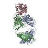
| ||||||||||
|---|---|---|---|---|---|---|---|---|---|---|---|
| 1 |
| ||||||||||
| Unit cell |
|
-Components
| #1: Antibody | Mass: 23583.869 Da / Num. of mol.: 1 / Fragment: light chain Source method: isolated from a genetically manipulated source Details: first chain of Anti-Lysozyme Antibody HyHEL-8 / Source: (gene. exp.)  |
|---|---|
| #2: Antibody | Mass: 22726.301 Da / Num. of mol.: 1 Source method: isolated from a genetically manipulated source Details: second chain of Anti-Lysozyme Antibody HyHEL-8 / Source: (gene. exp.) |
| #3: Protein | Mass: 14245.045 Da / Num. of mol.: 1 / Mutation: R61A Source method: isolated from a genetically manipulated source Source: (gene. exp.)  |
| #4: Chemical | ChemComp-ACY /   Mass: 60.052 Da / Num. of mol.: 1 / Source method: obtained synthetically / Formula: C2H4O2 Mass: 60.052 Da / Num. of mol.: 1 / Source method: obtained synthetically / Formula: C2H4O2 |
| #5: Water | ChemComp-HOH /  Mass: 18.015 Da / Num. of mol.: 546 / Source method: isolated from a natural source / Formula: H2O Mass: 18.015 Da / Num. of mol.: 546 / Source method: isolated from a natural source / Formula: H2O |
| Has protein modification | Y |
-Experimental details
-Experiment
| Experiment | Method: X-RAY DIFFRACTION / Number of used crystals: 1 |
|---|
- Sample preparation
| Crystal | Density Matthews: 2.25 Å3/Da / Density % sol: 44.8 % | |||||||||||||||||||||||||||||||||||
|---|---|---|---|---|---|---|---|---|---|---|---|---|---|---|---|---|---|---|---|---|---|---|---|---|---|---|---|---|---|---|---|---|---|---|---|---|
| Crystal grow | Method: vapor diffusion, hanging drop / Details: VAPOR DIFFUSION, HANGING DROP | |||||||||||||||||||||||||||||||||||
| Crystal grow | *PLUS Method: vapor diffusion, hanging drop | |||||||||||||||||||||||||||||||||||
| Components of the solutions | *PLUS
|
-Data collection
| Diffraction source | Source: SYNCHROTRON / Site: NSLS  / Beamline: X12B / Beamline: X12B |
|---|---|
| Detector | Detector: CCD |
| Radiation | Protocol: SINGLE WAVELENGTH / Monochromatic (M) / Laue (L): M / Scattering type: x-ray |
| Radiation wavelength | Relative weight: 1 |
| Reflection | Resolution: 1.9→33.32 Å / Num. all: 46457 / Num. obs: 46457 / Observed criterion σ(I): 0 / Biso Wilson estimate: 13.2 Å2 |
| Reflection | *PLUS Highest resolution: 1.9 Å / Num. obs: 46465 / % possible obs: 95 % / Num. measured all: 403172 / Rmerge(I) obs: 0.053 |
| Reflection shell | *PLUS Highest resolution: 1.8 Å / Lowest resolution: 1.9 Å / % possible obs: 80.7 % / Rmerge(I) obs: 0.3 / Mean I/σ(I) obs: 3.2 |
- Processing
| Software | Name: CNS / Version: 1 / Classification: refinement | ||||||||||||||||||||||||||||||||||||||||||||||||||||||||||||||||||||||||||||||||
|---|---|---|---|---|---|---|---|---|---|---|---|---|---|---|---|---|---|---|---|---|---|---|---|---|---|---|---|---|---|---|---|---|---|---|---|---|---|---|---|---|---|---|---|---|---|---|---|---|---|---|---|---|---|---|---|---|---|---|---|---|---|---|---|---|---|---|---|---|---|---|---|---|---|---|---|---|---|---|---|---|---|
| Refinement | Method to determine structure: MOLECULAR REPLACEMENT / Resolution: 1.9→33.32 Å / Rfactor Rfree error: 0.005 / Data cutoff high absF: 4249343.17 / Data cutoff low absF: 0 / Isotropic thermal model: RESTRAINED / Cross valid method: THROUGHOUT / σ(F): 0
| ||||||||||||||||||||||||||||||||||||||||||||||||||||||||||||||||||||||||||||||||
| Solvent computation | Solvent model: FLAT MODEL / Bsol: 49.1758 Å2 / ksol: 0.365696 e/Å3 | ||||||||||||||||||||||||||||||||||||||||||||||||||||||||||||||||||||||||||||||||
| Displacement parameters | Biso mean: 25.1 Å2
| ||||||||||||||||||||||||||||||||||||||||||||||||||||||||||||||||||||||||||||||||
| Refine analyze | Luzzati coordinate error free: 0.27 Å / Luzzati sigma a free: 0.23 Å | ||||||||||||||||||||||||||||||||||||||||||||||||||||||||||||||||||||||||||||||||
| Refinement step | Cycle: LAST / Resolution: 1.9→33.32 Å
| ||||||||||||||||||||||||||||||||||||||||||||||||||||||||||||||||||||||||||||||||
| Refine LS restraints |
| ||||||||||||||||||||||||||||||||||||||||||||||||||||||||||||||||||||||||||||||||
| LS refinement shell | Resolution: 1.9→2.02 Å / Rfactor Rfree error: 0.016 / Total num. of bins used: 6
| ||||||||||||||||||||||||||||||||||||||||||||||||||||||||||||||||||||||||||||||||
| Xplor file |
| ||||||||||||||||||||||||||||||||||||||||||||||||||||||||||||||||||||||||||||||||
| Refinement | *PLUS Highest resolution: 1.9 Å / Lowest resolution: 100 Å / % reflection Rfree: 5 % / Rfactor Rfree: 0.254 / Rfactor Rwork: 0.215 | ||||||||||||||||||||||||||||||||||||||||||||||||||||||||||||||||||||||||||||||||
| Solvent computation | *PLUS | ||||||||||||||||||||||||||||||||||||||||||||||||||||||||||||||||||||||||||||||||
| Displacement parameters | *PLUS | ||||||||||||||||||||||||||||||||||||||||||||||||||||||||||||||||||||||||||||||||
| Refine LS restraints | *PLUS
|
